When light propagates through a disordered medium, it gets scattered numerous times, which scrambles the optical wavefront. This is why biological tissue, bone, colloidal suspension, clouds, and fogs are opaque. Despite its complexity, this multiple scattering process is reversible thanks to the time-reversal symmetry of Maxwell’s equations. One goal of the Optics in Complex Systems group is to leverage this property and “unscramble” light scattering to see the parts of the physical world blocked by opacity.
By time-reversing the multiply-scattered light emitted from a point source (i.e., a guidestar) inside a scattering medium, one can focus light back to the originating source location. Scanning that source location can, in principle, create a 3D image of the medium. However, finding the appropriate wavefront used to require physical guidestars or detectors at the target locations, which come with many limitations such as being invasive, requiring fluorescence labeling, degrading the resolution, limited within a small isoplanatic patch, restricted to planar targets outside the scattering media, and slow wavefront updates due to the hardware.
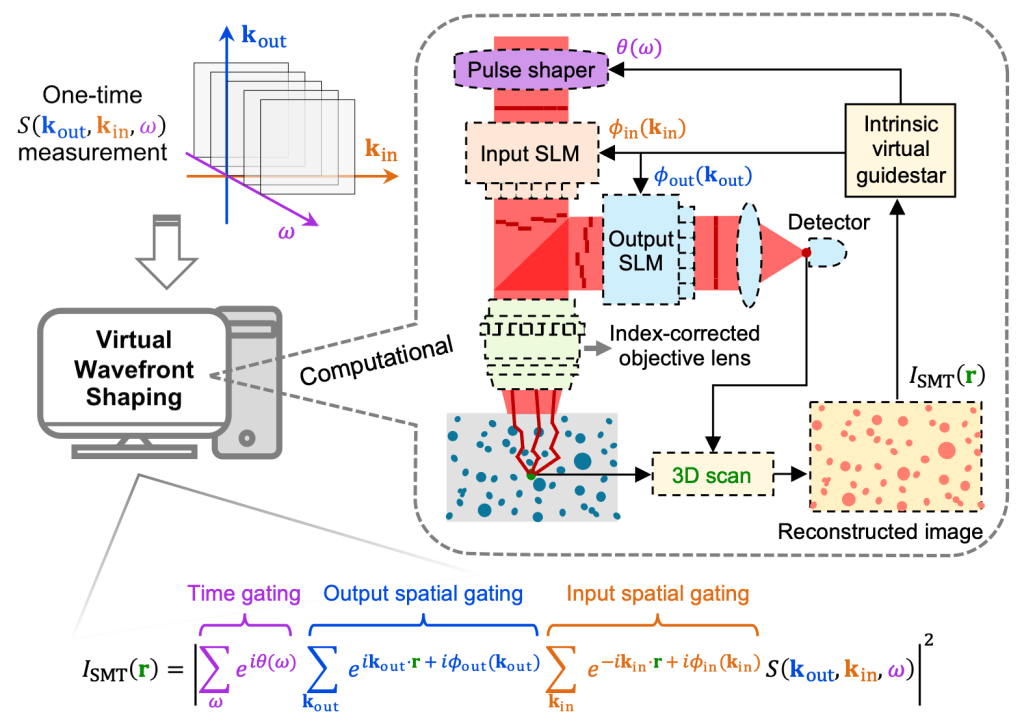
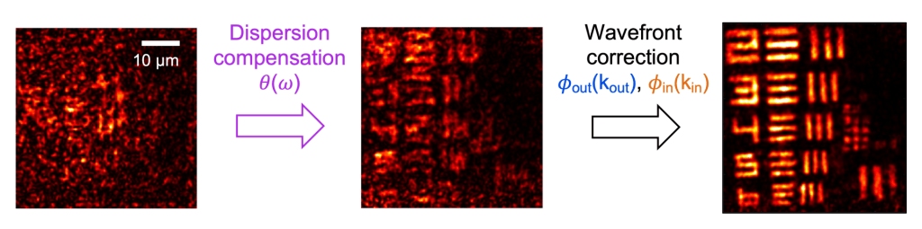
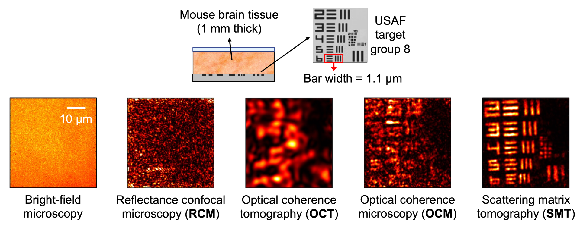
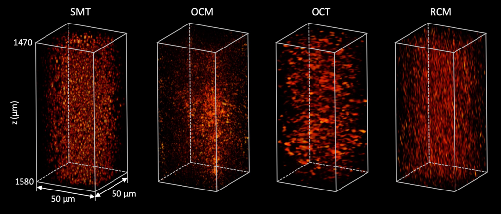
We develop an approach called “scattering matrix tomography” (SMT) [1,2] that converts the problem of imaging and reversing light scattering to a computational reconstruction and optimization problem, as illustrated in the figure below. The scattering matrix element S(kout, kin, ω) is the outgoing amplitude in direction kout given an incident wave from direction kin at frequency ω. We measure the scattering matrix of the sample, use it to digitally scan a synthesized confocal spatiotemporal focus and construct a coarse volumetric image of the sample, and then use the synthesized image as many virtual guidestars to digitally optimize the dispersion compensation θ(ω), input wavefront ϕin(kin), and output wavefront ϕout(kout) to compensate for aberrations and scattering. The virtual feedback dispenses with physical guidestars and enables hardware-free spatiotemporal wavefront corrections across arbitrarily many isoplanatic patches. SMT is noninvasive and label-free, and it provides volumetric images inside and outside the scattering media with unprecedented depth-over-resolution ratios. It can be applied to medical imaging, device inspection, biological science, and colloidal physics.


Related publications
- Deep imaging inside scattering media through virtual spatiotemporal wavefront shaping, Yiwen Zhang, Minh Dinh, Zeyu Wang, Tianhao Zhang, Tianhang Chen, and Chia Wei Hsu. arXiv:2306.08793.
- Full-wave simulations of tomographic optical imaging inside scattering media, Zeyu Wang, Yiwen Zhang, and Chia Wei Hsu. arXiv:2308.07244.