
Research Overview
The Shahidi Laboratory is focused on development and application of prototype optical imaging technologies to study pathophysiology and improve diagnostic evaluation of ocular and systemic diseases. Ongoing research projects include: imaging of retinal oxygen delivery and metabolism in experimental models of retinal diseases, characterization of microvascular hemodynamics in ocular and systemic diseases, identification of image-based biomarkers of diabetic retinopathy, and assessment of retinal neurovascular coupling and oxygen metabolism in diabetic retinopathy, sickle cell retinopathy and glaucoma.
Research Topics
- Ocular Imaging Technology
- Ocular Hemodynamics
- Neurovascular Coupling
- Retinal Oxygen Metabolism
- Image-based Biomarkers of Disease
Ongoing Projects
Project 1: Imaging of Retinal Oxygenation and MetabolismProject 2: Ocular Imaging Biomarkers of Diabetic RetinopathyProject 3: Identifying Oxygen-related Ocular Biomarkers of GlaucomaProject 4: Relating Tissue Perfusion and Oxygenation in Retinal Diseases
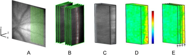
Project 1: Imaging of Retinal Oxygenation and Metabolism
Development of multimodal imaging techniques that measure retinal vascular/tissue oxygen tension and blood flow, and assess retinal oxygen delivery, metabolism, and extraction fraction under experimental ischemic conditions.
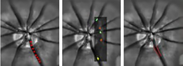
Combined imaging of retinal tissue and vascular oxygen tension in rat using dual oxyphor optical section phosphorescence lifetime imaging
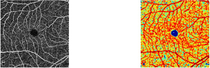
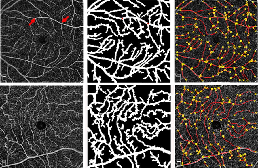
Project 2: Ocular Imaging Biomarkers of Diabetic Retinopathy
Identifying ocular biomarkers of microvascular, neural, and metabolic function that are predictive of development, progression, and treatment outcome of diabetic retinopathy.
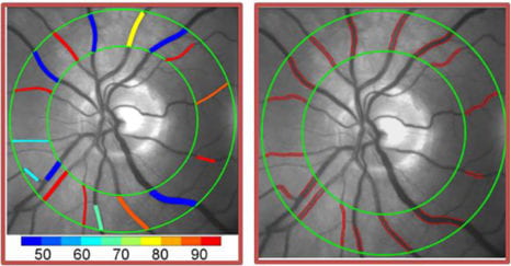


Project 3: Identifying Oxygen-related Ocular Biomarkers of Glaucoma
Combining oxygen-related metrics that have been individually proven to be disturbed in glaucoma to identify novel oxygen-related biomarkers that directly and comprehensively assess retinal vascular and functional reserve for predicting development, progression, and treatment of glaucoma.
Project 4: Relating Tissue Perfusion and Oxygenation in Retinal Diseases
Funding
- NIH/NEI- R01: Imaging of Retinal Oygenation and Metabolism
- NIH/NIDDK- DP3: Ocular Biomarkers of Microvascular, Neural and Metabolic Function in Diabetes