![]()
Yeu Q Nguyen & Mariam Davtyan, PhD, MPH & Toni Frederick, PhD, MSPH
Kristine Augustyn & Eric J. Kezirian, MD, MPH
Ching Ching Cheng & Raymond L. Goldsworthy, PhD
Ester Petschar & W. Martin Kast, PhD
Karen Ruth Karlsson & Evanthia Roussos Torres, MD, PhD
Billy Pacak & Leonardo Morsut, MD, PhD
Cathy Immordino & Zhanghua Chen, PhD
Rebecca Niederlander & Axel Montagne, PhD
Larissa Nickel & Shuwan Liu, PhD Candidate
Susan Price & Yuli Talyansky, PhD Candidate
Ella Mikayelyan & Sheila Pakdaman, MS, PhD Student Research Assistant
Lisa Lesniak & Ya-Wen Chen, PhD
Ted Meyer, Curator
What happens when you pair Los Angeles’s most creative artists with Keck School of Medicine’s world-class researchers? The HEAL Program’s 4th annual Artist and Researcher Show!
The Artist and Researcher exhibit series highlights the parallel processes of scientific curiosity and artistic inquiry with the aim of making complex research more understandable to the general viewing public. Each artist learns about the work of his or her researcher through dialogue and/or lab visits, and creates a visual language to make the science more accessible. Over the past four years, the Artist and Researcher exhibits have paired 47 teams with stunning results.
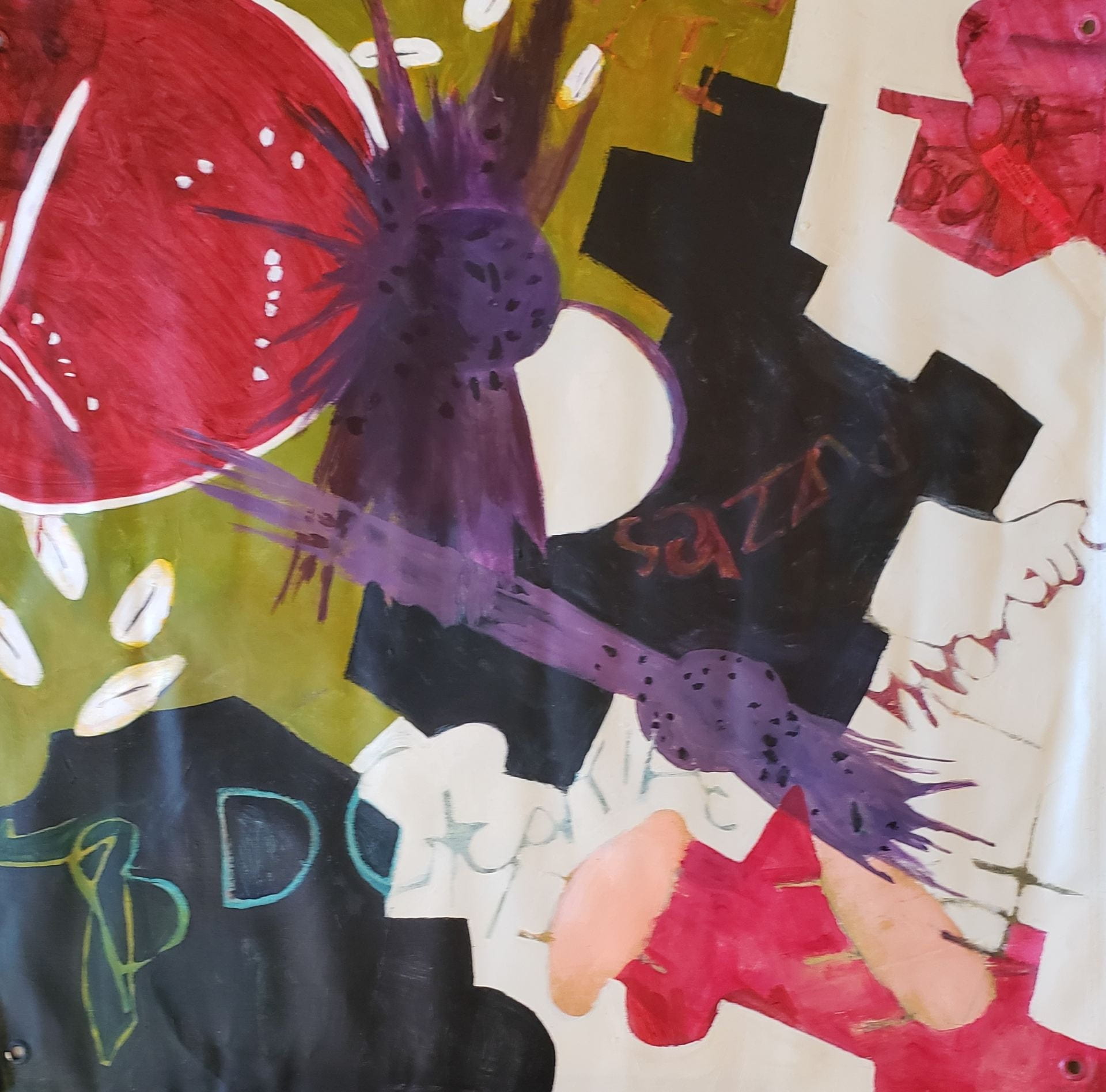
Reframe, Rework, Revive
Series of digital photography and collage painting
by Yeu Q Nguyen

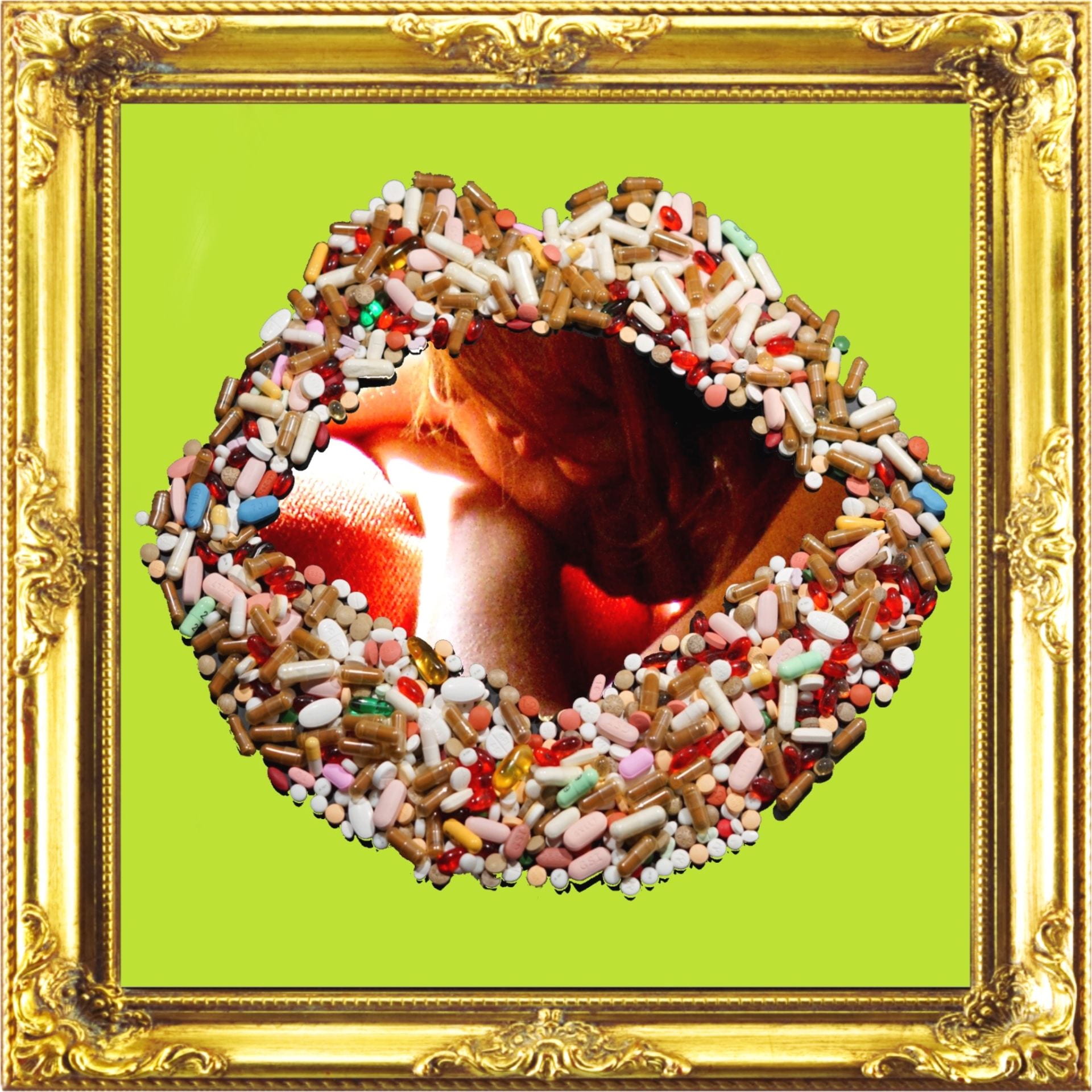
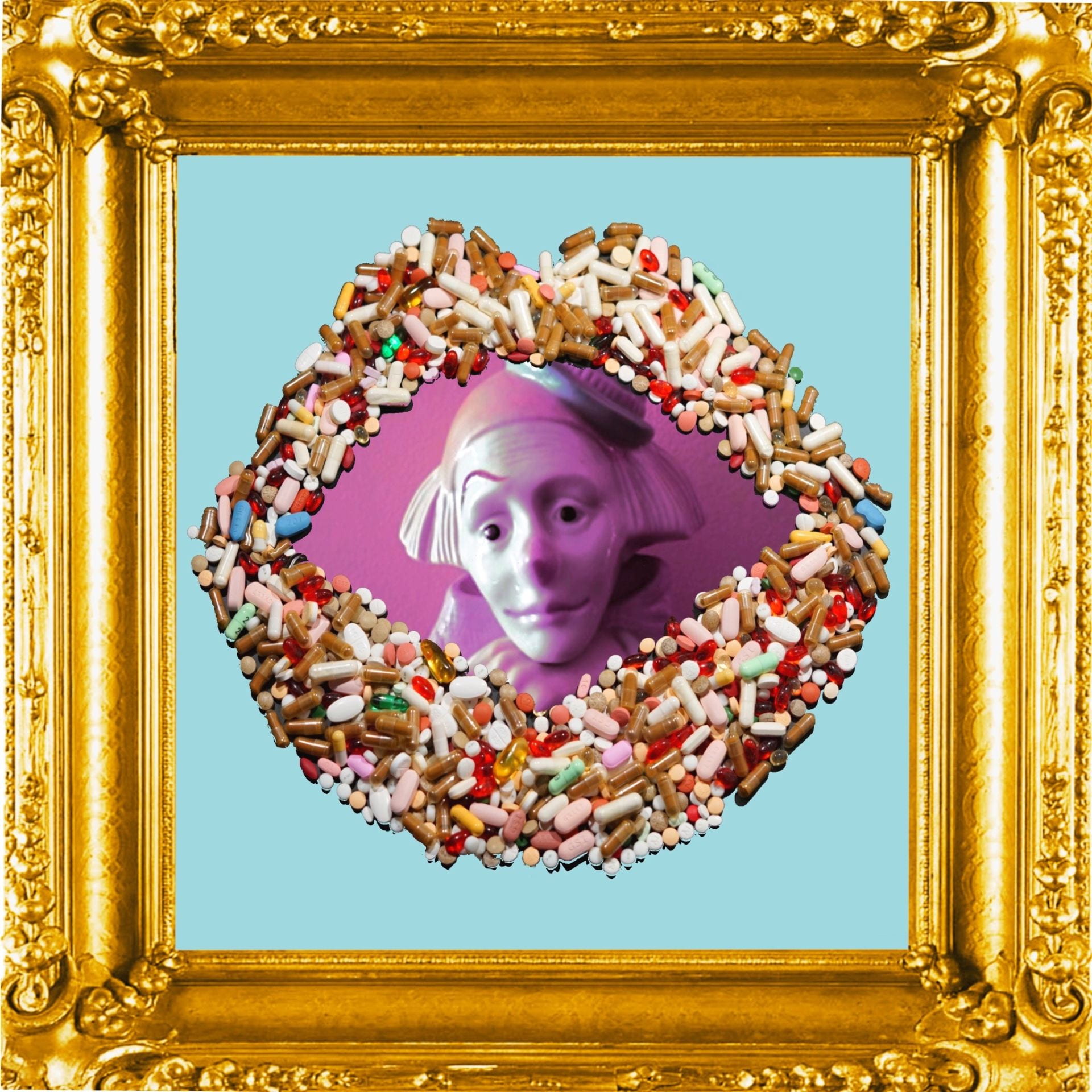
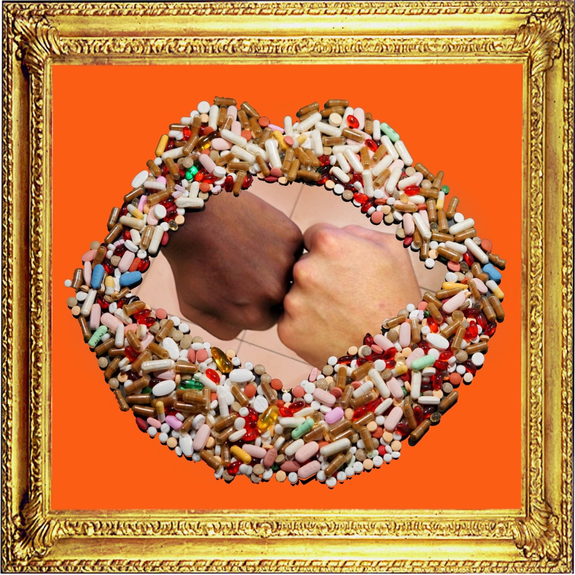
Adding eye-popping, look-at-me colors to the photographs submitted by anonymous HIV-positive patients who participated in the Adherence Treatment with Photo Stories (AAPS) clinical study, I rework private records of pain, heartbreak, and suffering into trendy, iconic images representing human resilience and courage to live in the face of chronic illness and HIV-related stigma. These intimate stories, which are often relegated to hushed tones and locked rooms, deserve more public attention as well as admiration. The world is but a mirror we hold up to see ourselves. We might not be able to change how the mirror reflects our image, but we can always choose how we want to see our own reflection.
The average HIV-positive patient consumes about 155,000 pills in their entire lifetime, not counting additional vitamins, supplements, or pills treating other ailments that naturally occur during our lifespan. These pills must also be taken everyday, at the exact same time, without fail. This is a monumental task, requiring absolute discipline and commitment to LIFE. Reframing their stories in beautiful, ornate, and precious gold frames, I want to celebrate these extraordinary women, who have chosen to live, love, and revive in the face of illness and despair.
Website: www.yeuqart.com
Instagram: @yeuqnguyen
Addressing Adherence with Photo Stories (AAPS)
Mariam Davtyan, PhD, MPH & Toni Frederick, PhD, MSPH
HIV (human immunodeficiency virus) is a virus that attacks the body's immune system. If HIV is not treated, it can lead to AIDS (acquired immunodeficiency syndrome). There is currently no effective cure. Once people get HIV, they have it for life. However, HIV can now be controlled by strict adherence to anti-retroviral therapy (ART) which can dramatically slow the disease's progress, prevent secondary infections and complications, and prolong life. Today, HIV is a chronic and manageable condition. Despite so many advances in treating and normalizing life with HIV, what has not changed is the stigma attached to living with HIV. Stigma refers to exclusionary attitudes and behaviors directed towards people living with HIV and those presumed by others to be living with HIV. Nearly 90% of HIV patients report being stigmatized due to their disease status, and the human impact of this stigma can be devastating. HIV stigma is associated with poor self-regard, self-blame and shame, reduced adherence to life-saving ARTs, and mental health problems, including stress, depression and suicidal ideation. Stigma can be one of the most damaging symptoms of HIV: It destructs, disrupts, and disempowers. The Maternal, Child and Adolescent Center for Infectious Diseases and Virology (MCA Center) at LAC+USC, is a state-of-the-art family-centered model of care combining primary care, tertiary care and research for disenfranchised and traditionally hard-to-reach HIV+ populations. MCA Center’s unique comprehensive one-stop model of care enables women, men, children and adolescents to receive a continuum of primary care with specialty care services including medical evaluation and clinical care, psychosocial support services, adherence counseling, and nutritional counselling. Little is known about how this group of disenfranchised and hard-to-reach HIV+ persons perceive their lived experiences with HIV stigma and how this stigma may be impacting their engagement in care. To understand more about the impact of HIV-related stigma on this population, we conducted a PhotoVoice-informed research study. PhotoVoice is a community-based participatory research methodology rooted in feminist theory, empowerment education, and documentary photography. Study participants were recruited from the MCA Center, trained on the concept of PhotoVoice and asked to take photos reflecting their experiences with HIV stigma. They were asked to use the next few weeks to take their photos and write a caption describing how their photo captured their feelings of HIV-related stigma. We then asked them to return for a debrief session to discuss their photographs in a group setting with our MCA pharmacist. The debrief sessions revealed that participants felt an overwhelming sense of shame and guilt for having HIV. They self-identified as “dirty,” “contagious,” and “marked.” However, participation in the process of taking photographs to describe their experiences of HIV stigma, was found to be therapeutic. Some participants began to feel better about living with HIV and acquired a more positive thought process about the disease, while others felt the need to advocate and assist other patients in similar circumstances. A few participants expressed that they were compelled to improve their adherence to antiretroviral medications. Yeu has showcased 4 photos from this intervention. Below are the women’s captions for these photos:- "Contagious!"
- "Isolation, loss of life, and HIV!"
- "Sad clown…I can make everyone else happy, but I'm not happy!"
- "It's a worldwide fight: Black or White, HIV does not discriminate!"
To Sleep, Perchance to Dream
Acrylic on wood panel
by Kristine Augustyn
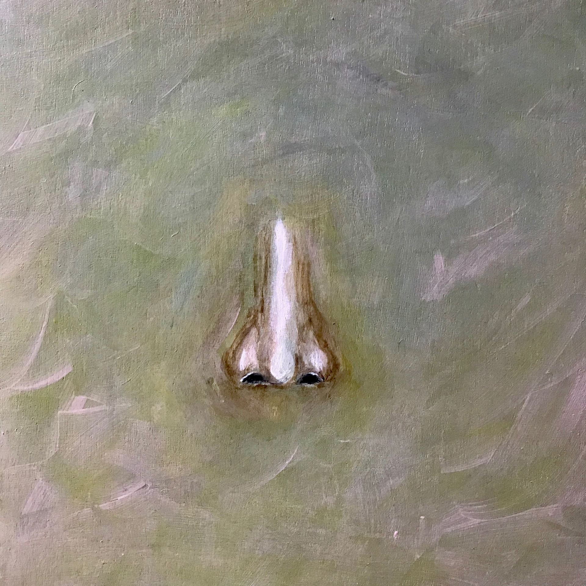

These three pieces were based on the work of Dr. Eric Kezirian, who specializes in the surgical evaluation and treatment of snoring and obstructive sleep apnea.
My inspiration was based on the endoscopic exploration Dr. Kezirian does to pinpoint and accurately identify each individual’s specific needs to help with their chronic sleep issues.
Sleep is such an important part of life, a time to revive and regenerate. I believe dreaming is also as important and as an artist I would be lost without my dreams.
Website: www.kristineaugustyn.com
Instagram: @kristineaugustyn
Twitter: @kna17
Drug-Induced Sleep Endoscopy in Obstructive Sleep Apnea
Obstructive sleep apnea (OSA) affects over 20 million American adults and an estimated 900 million adults worldwide. OSA is defined by blockage of breathing during sleep, and OSA leads not only to disruptive snoring but also to sleep disturbances (resulting in poor sleep quality) and health effects (high blood pressure, heart attack, stroke, and death). Treatments include conservative approaches (weight loss, sleeping on one’s side, and avoiding alcohol within 3 hours of bedtime), positive airway pressure therapy (for example, CPAP), surgery, and oral appliance mouthpieces. In addition to using conservative measures, OSA patients who do not tolerate first-line treatment (positive airway pressure) may consider surgery.
Surgery is designed to open the space for breathing, and there are multiple structures of the throat that can contribute to blockage in breathing (such as the soft palate and/or tongue). Our research has contributed to major advances in OSA surgery over the past 15-20 years, but still only about 60-70% of patients get the results we want to achieve. There is clearly room for improvement.
Accurate identification of the anatomic cause(s) of OSA is essential. Drug-induced sleep endoscopy is an evaluation procedure that determines the structure(s) that contribute to OSA in a specific patient. Drug-induced sleep endoscopy involves administering sedation to reproduce conditions somewhat similar to natural sleep so that a surgeon can examine the throat with a flexible fiberoptic telescope and visualize the blockage in breathing directly. This allows use to determine what structures are causing OSA, with the goal of developing a tailored, effective treatment plan.
With European colleagues, we have developed the VOTE Classification, the standard way to characterize drug-induced sleep endoscopy findings. We have also led international multicenter studies showing how drug-induced sleep endoscopy can identify patients who may do better and worse with various procedures. We are excited about the work to date and the many important, unanswered questions that remain!
Absent of Stimulation
Cinematography
by Ching Ching Cheng
Password: chinguscproject
This video work is exploring the relationships between auditory, brain, and memory. When we hold a seashell up to your ear, we can hear the waves of the ocean, as if the sounds from the seashell’s past environment are still echoing within it. But what we actually hear is the sound of resonating air produced by the shell’s cavity. Whenever there are missing sounds, the brain creates imaginary sounds to fill the gap. When we can not find answers from science, instead, we tend to look for spiritual meanings. Scientifically, ringing in the ears could be one of the symptoms of hearing loss, but many people also believe that It is the moment of spiritual awakening. The sound or sensation of ear ringing is due to being tuned into the divine sound of the universe and resonating with source energy.
A video by Ching Ching Cheng
Cinematographer: Aakash Raj
Email: kinoko477@gmail.com
Website: chingchingcheng.com
Instagram: @chingchingcheng
I am a man not a tree
Acrylic and fluorescent paint
by Ester Petschar
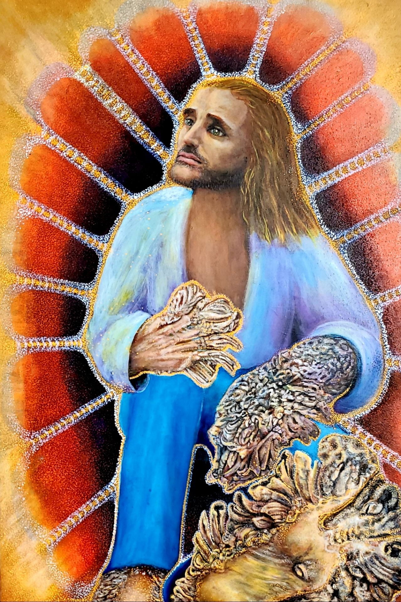
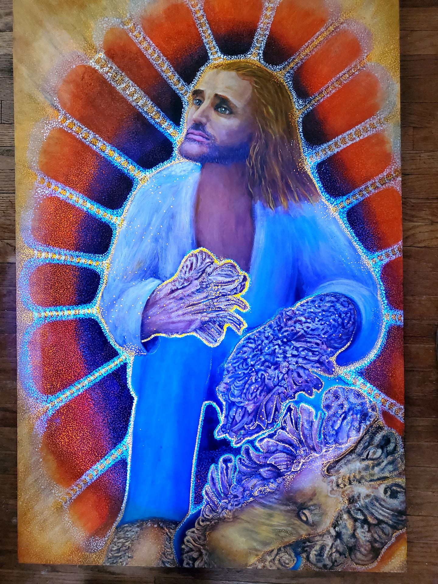
This piece conveys Dr. Kast's work in a simple way for the viewer to see what happens to a person with this disease. The blue surrounding the normal red cell, is being penetrated by the gold color in the body of a man (Dede Kasawara). The fluorescent colors are enhanced by black light. The gold represents a rare disease which causes a genetic variation resulting in the tree-bark like warts.
Email: dotartla@gmail.com
Instagram: @esterpetschar
A few years passed and Discovery Channel visited the patient again. All the warts had regrown with a vengeance. As Discovery Channel knew that warts were caused by some human papilloma virus (HPV) strains and hypothesized that an immune deficiency was likely preventing the patient from mounting an immune response against HPV, they approached Dr. W. Martin Kast, a renowned viral immunologist at USC, for advice. This led to Dr. Kast’s expedition into the jungle of Indonesia and a third documentary called “Treeman Meets Treeman” After Dr. Kast met Dede, he subsequently identified two more patients. Zainal in Indonesia and Ivan, born in the Netherlands, who turned out to be of Indonesian decent. The documentary reveals that all three Treeman had family ties to a small village near a lake near Bandung in West Java and therefore were likely related. In the documentary arrangements were made for the two Indonesian patients to meet each other, explaining the title of the third documentary.
There are some rare reports in the medical literature about patients with a similar disease called Epidermodysplasia Verricuformis, a disease caused by infection with HPV types 5 or 8 and involving mutations in the EVER 1 or 2 genes. Dr. Kast has since identified two more Treemen and analyzed the tissue samples of all five patients. They all have mutations that effect skin immunity. They were all infected with HPV, but their types were HPV 2, 27 or 57 and none had mutations in the EVER 1 or 2 genes. Research is continuing on these patients but it is clear that this is a newly identified rare disease. Dr. Kast’s personal interactions with these patients show that, despite their frightening appearances, these patients are human beings. They are men not trees. The artist Ester Petschar aimed to exemplify that in this painting in which she also depicted how “the red orange cells of Dede are being invaded by the golden bad virus.” This work is a tribute to the lives of Dede and Ivan, who have unfortunately died. But their suffering might not have been in vain as it could yield information on why patients that have other HPV related diseases, like cervical cancer, do not clear HPV. May these Treemen rest in peace.
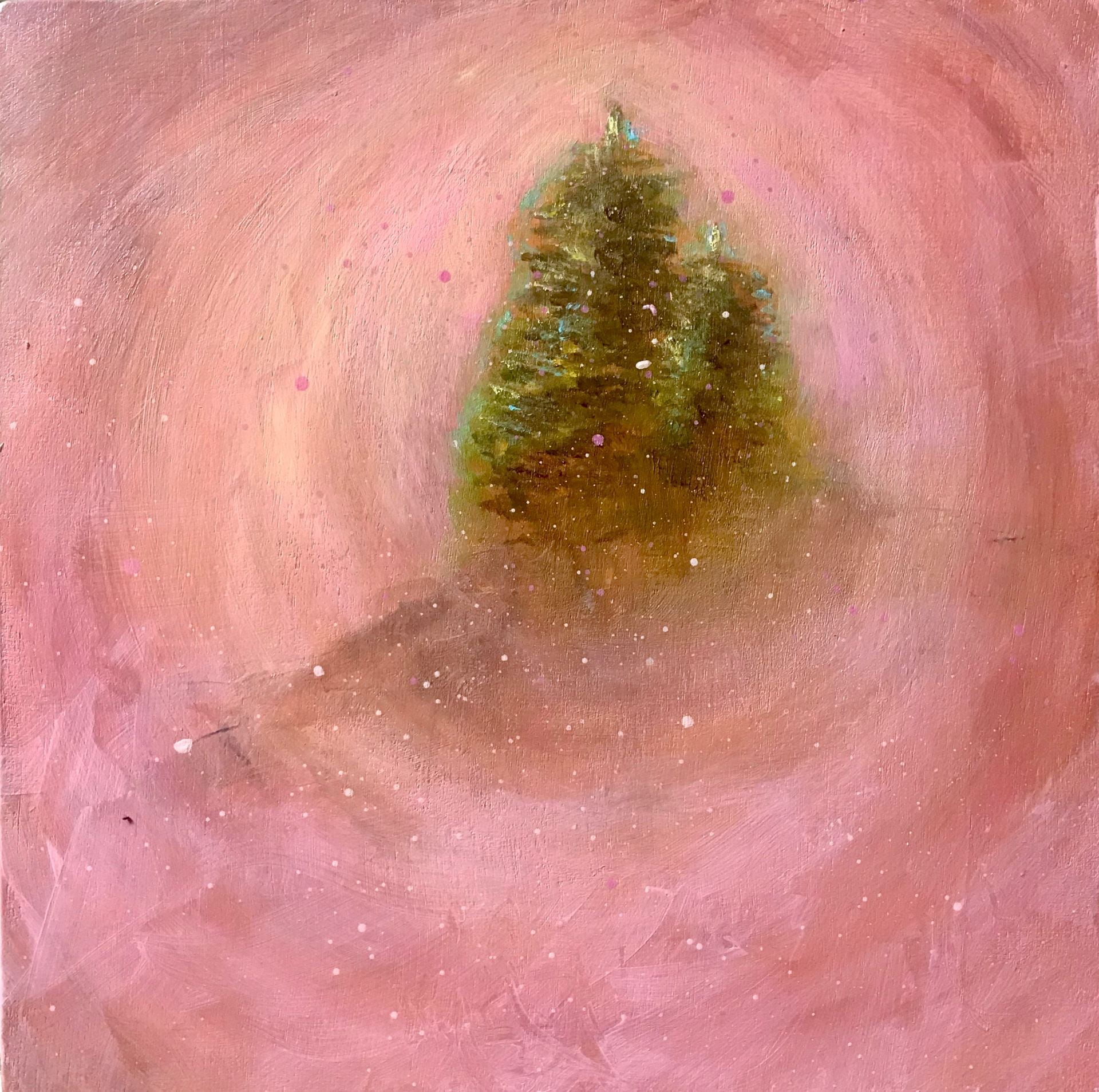
Heat Wave
Encaustic and oil on panel
by Karen Ruth Karlsson
Dr. Roussos-Torres’ research is focused on identifying ways to change the environment inside breast cancer tumors to make the tumors more sensitive to immunotherapy. She described the goal as turning “cold” tumors full of immune-suppressing cells into “hot” tumors full of T-cells and other cancer fighters. “Heat Wave” depicts a cold tumor environment becoming hot after the right pathway is found. This piece was created in encaustic and oil. Encaustic paint is made up of beeswax, resin and pigment which must be melted by the application of heat to render it useful for painting.
E-mail: karlssonarts@gmail.com
Website: www.karlssonarts.com
Instagram: @karlssonarts
Facebook: KarlssonArts
Sculpture, Untitled
by Bill Pacak
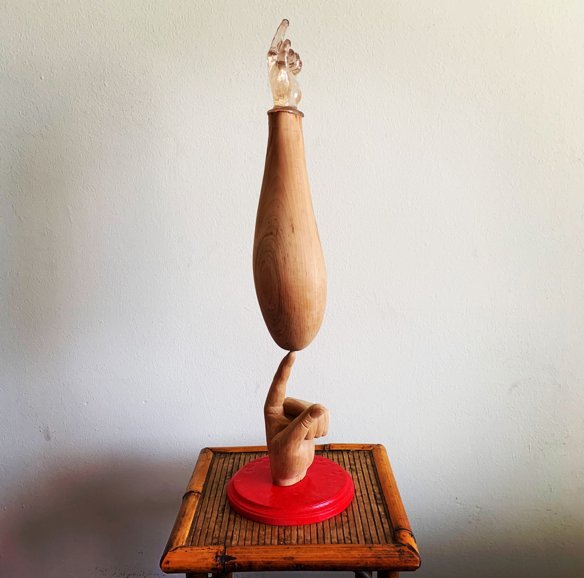
The idea of genetically modified stem cells to regrow peoples limbs and organs is a pretty ripe area for an artist to take on. Leonardo’s research is extremely exciting. I was quite surprised to find out that he never watched the Deadpool movie. This is the technology that could make superheroes, let alone save peoples lives by growing them new hearts and limbs. With his peace I took a very literal interpretation in a surrealist way on how this technology could look.
Email: billpacak@gmail.com
Instagram: @billy_pacak
In our lab we start from the realization that embryonic development is a technology for building tissues, based on genetically encoded networks of signaling and differentiation. In an attempt to master this technology to build designer, smart tissues, of user-defined structure and function, we invent and use synthetic biology tools to control multicellular behaviors, with a special focus on stem cells and regenerative medicine applications. One day, we are hoping to be able to learn to control the organ building technology so well to be able to grow back entire organs or body parts, which we imagine will be comprised of part normal, part synthetic, designer cells.
Website: http://morsutlab.usc.edu/
Website: https://sites.usc.edu/cieborg/
Twitter: @LeonardoMorsut
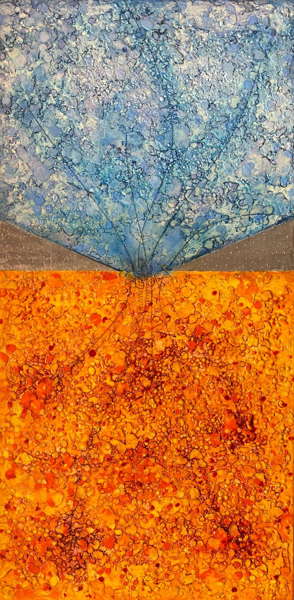
Metabolomics
Archival pigment print
by Cathy Immordino
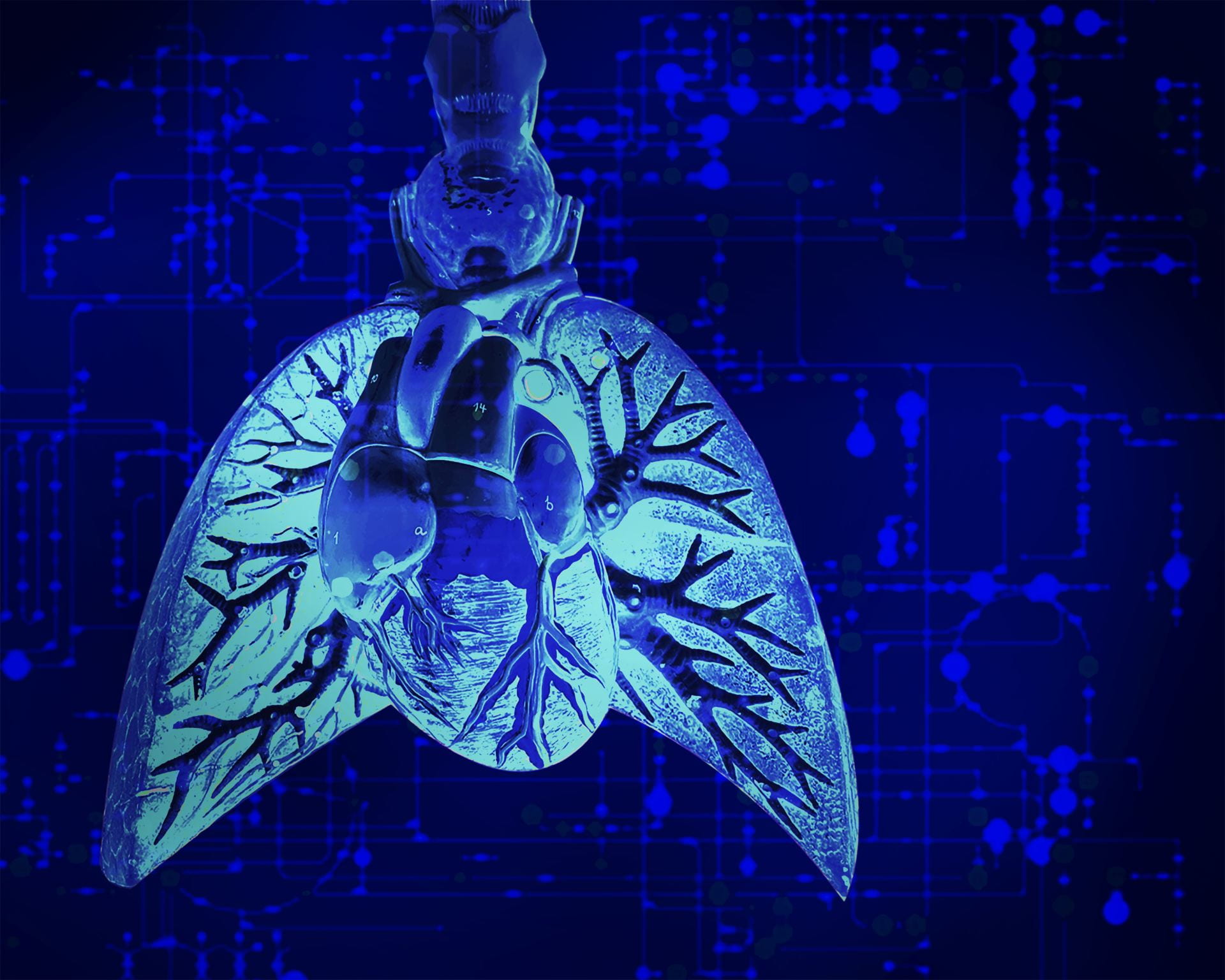
Metabolomics – how one's body processes everything surrounding them, their environment, the air they breath, the food and water their ingest. Molecules enter the lungs, are processed, and affect the wellbeing of the person.
Inspired by the research of Zhanghua Chen, I created this piece to represent one of the main talking points from our encounter – that air quality and pollution is linked to disease. I set out to make a simple collage. Lungs were captured, colors over-saturated and heightened to show toxicity like you would see in a poison dart frog. Behind, part of the metabolomic chart rests amongst light and dark areas suggesting inconsistencies in the environment. The environment enters the body in many ways, especially through the throat on the way to the lungs. The blue represents the throat chakra in this regard and how the lesser understood environmental factors contribute to the toxicity of one' health.
Website: http://www.cathyimmordino.com
Facebook: https://www.facebook.com/cathyimmordinoart
Instagram: https://www.instagram.com/cathyimmordinoart
Twitter: https://twitter.com/cathyimmordino_
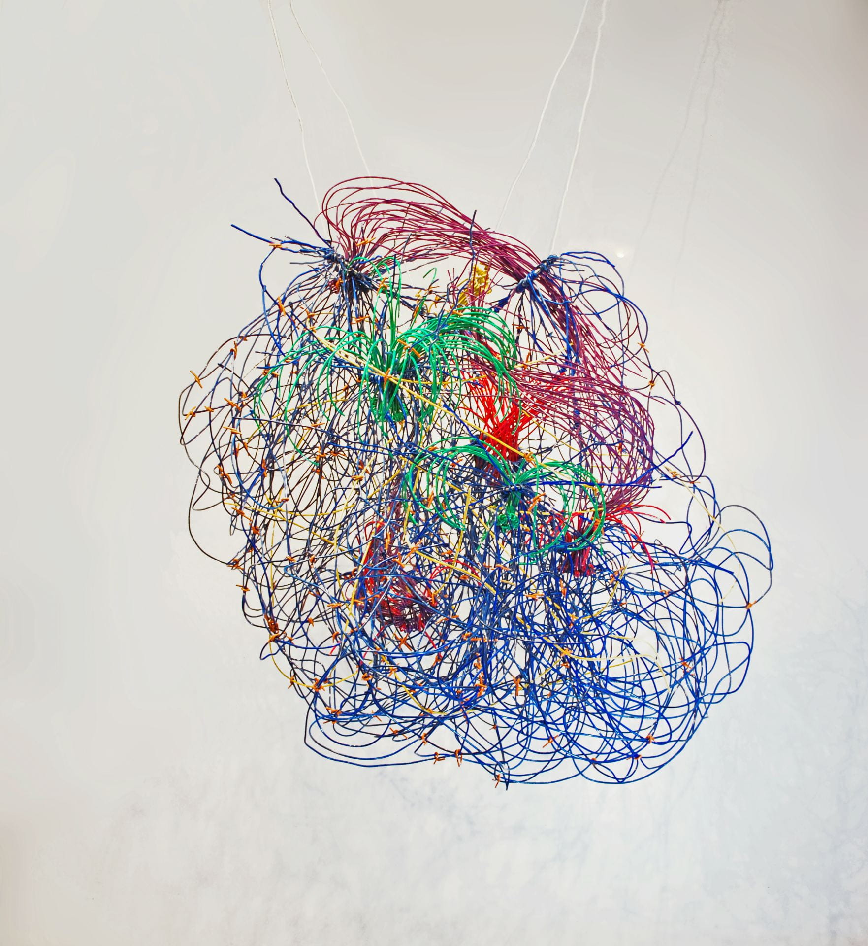
A Bundle of Matters
Plastic coated copper conduit
by Rebecca Niederlander
Community. Connection. Responsibility.
My practice is complicated individual elements brought together precariously; how they connect together, stressed by gravity and tension. Responsible for each other, their precarious nature mirrors their own mortality and their responses to the stressors of existence mimics our own. I love making suspended works; they defy what they can, rebellious imps that they are, while having to acknowledge what they must.
I am a materialist in the truest sense of the word. I interrogate my materials lovingly and with great care looking for the ways in which they can speak to commonalities or show their uniqueness. And what of this compulsive need for connection? I stand there, with you, be it in person or in spirit through the work experiencing the micro and macro of existence; the before and now and future and wondering why we are crossing paths. For while I know that I am a spec of dust, as are you, it is stardust. Every atom in our bodies came from a star that exploded. We are in the middle of our bodies, our families, our communities, the Milky Way.
At its essence: Things fall apart and reconnect. I document it.
How wonderful then, to be paired by Ted Meyer with Dr. Axel Montagne. His research into the ways bits of our body are responsible for the other bits is such a poignant microcosm of the entirety of, well, everything. And he, too, documents what falls apart, but he tries to figure out the why and is having some amazing successes. In the meantime he makes some gorgeous imagery that stimulated me to produce a playful suspended wire sculpture simulacra of the brain wirings.
We have had some wonderful conversations, he and I. And for those I am grateful. Both from the perspective of an artist learning more about scientific things I enjoy, certainly. But more so because as a person with an inflammatory chronic illness Dr. Montagne has been illuminating in helping me to understand some of the things people like me experience.
Wesite: www.becster.org
Instagram: @rebeccaniederlander
My research training has provided me with comprehensive and diverse background in a wide variety of scientific disciplines including molecular and cellular biology, magnetic resonance imaging (MRI), translational research, and the pathophysiology of neurodegenerative diseases such as Alzheimer’s disease (AD) and related dementias as well as ischemic and hemorrhagic strokes. Specifically, my true passion is in the area of neuroscience, particularly the research to understand biological processes that cause cerebrovascular disorders and ultimately lead to cognitive decline; this is an increasingly important avenue of research as the world’s aged population continues to rise. The potential to improve diagnosis using novel neuroimaging methods and to find new treatments for dementia and/or stroke patients combined with the intellectual challenge of basic research is both rewarding and motivating.
During my young research career, I’ve acquired a strong arsenal of skills particularly in brain imaging methods including advanced microscopic imaging and MRI of cerebrovascular inflammation, neurovascular dysfunctions, and neuronal connectivity in both pre-clinical animal models and clinical datasets. I’ve published several major findings in high-profile journals such as Nature, Nature Medicine, Nature Neuroscience, Neuron, and so on. I believe that my most important findings have already impacted our research field with new imaging techniques that are now available worldwide as well as a better knowledge of how vascular dysfunctions contribute to neuron loss and dementia. In 2012, I developed molecular MRI of neuroinflammation using iron oxide particles that target inflamed vessels. This approach exhibits clinical potential to identify and select patients that would be most likely to benefit from anti-inflammatory treatment in several neuropathological conditions including AD and stroke. In 2015, I developed an advanced MRI method allowing detection of subtle vascular leakages in the living human brain. I’ve shown that subtle vascular breakdown during normal aging begins in the hippocampus, a region critical for learning and memory. In a 2019 follow-up study, I also demonstrated that an accelerated vascular breakdown may contribute to cognitive impairment, independently of the classical AD amyloid and tau biomarkers. In 2018, I used novel cutting-edge imaging methods to show that capillary breakdown initiates white matter (WM) dysfunction in the brain. Importantly, I’ve found that targeting blood-derived fibrinogen and limiting these protein deposits in the brain can slow WM disease, which has implications for the pathogenesis and treatment of human WM disease associated with AD. In 2019, I showed that brain capillary disruption leads to rapid circulatory failure and neuronal loss via the loss of a specific capillary growth factor called pleiotrophin. Finally, in 2020, I’ve found that people carrying the APOE4 gene, the major genetic risk for AD, have subtle cerebrovascular leakages which can be detected years prior the build-up of amyloid and tau in the brain and years prior cognitive decline. I’ve also discovered the cellular and molecular mechanisms explaining this increased brain vessel permeability in people at risk for AD. The good news is that the biological pathway is targetable, and we are currently working on drug candidates that could seal the leaky blood vessels which would ultimately protect neurons and preserve cognitive functions.
Neuronal Connectivity: I was also involved in developing a new hybrid diffusion imaging (HYDI)-derived diffusion tensor imaging (DTI) method that may be valuable in human connectome projects and clinical research, as well as MR research in experimental animals. This method allows precise post-processing connectivity data with powerful tract tracing of white matter fibers in both humans and animal models. I’ve been using ultra-high field 11.7 Tesla MRI for animals and clinical 3 Tesla MRI in humans. I’ve worked on high-resolution tractography maps allowing the detection of different white matter circuitry (e.g., cortico-callosal, corticospinal, and corticolimbic pathways) running through the brain. Lastly, I’m currently analyzing DTI metrics in different brain subregions of AD and mild dementia patients scanned at USC and Washington University, and trying to bring out interesting correlations with the MRI vascular measures as well as cerebrospinal fluid (CSF)/blood biomarkers and cognitive functions.
Email: montagne@usc.edu
LinkedIn: https://www.linkedin.com/in/axelmontagne
Twitter: @AxL_Montagne
Periosteal Bone Regeneration
Acrylic paint, fabric, lace, and beads
by Larissa Nickel
"It's what's happening this time, so I'm thankful for letting me see another spring."– Nina Simone, Another Spring, 1969.
Our bodies are an architecture of existence. Periosteal Bone Regeneration utilizes form, materiality, and symbolism to denote the visual physical layers of the body, and the related conceptual signification of stem cell bone regeneration research. Feminist craft materials of lace, fabric, and textiles recreate the microscopic cellular view of scientific investigations of the body. In part a diagrammatic view of bone, periosteal cells, and skin rendered as materialism, the work also imitates "nature in her manner of operation" (John Cage) through the ideology of spring as renewal.
The ecological definition of spring relates to its biological indicators such as blooming flowers and flora, soil temperature, and animal activities. In the Transformation of Nature in Art, Ananda Coomaraswamy interprets spring as regeneration using Indian philosophy and symbolic meaning. Shuwan Liu's USC research on the periosteum–the connective tissue covering the external surface of bone–and discovery of a unique periosteal cell (Sox 9+), intends to contribute to bone regenerative processes. Together our practices intersect art, science, technology, philosophy, and narrative to explore the worlds of creative practice, object and concept, and the connection that collectively, no matter what's happening this time, we will see a regenerated spring.
Email: lnickel1@alumni.jh.edu
Website: larissanickel.com
Instagram: @larissa.nickel
a baumanni, on the run
Acrylic and USC Medical ID band on canvas
by Susan C. Price
I visited Yuli in his lab. This was convenient as I visit Norris Cancer Center monthly as a lab rat in a large, international study related to Alzheimer’s. Yuli explained what he was doing, partly by showing me a creative, colorful power point he had made. Then Yuli and his fiancé visited my home studio. It has been a great pleasure to get to know a scientist.
Science is beautiful, and critical to our survival. I admire Yuli’s love of the puzzle. I view trying to make a painting “work”, as a puzzle. What colors/shapes “play nice” with the others? I try to achieve an off-balance balance of large to small, dark to light.
I started this work by painting words and phrases relating to the bacteria, and some depictions of the bacteria from the power point Yuli gave me. The words include: “pneumonia”, “puzzle”, “sepsis”, “no tools left”, “in love with science”. I painted over most of that, and turned the canvas upside down. Sometimes painting over the words leaves parts visible, which often become pattern. Or, they don’t show at all, except in my memory. A bit mystical, but it entertains me.
Acinobacter baumannii is a strange, wonderful and tricky/horrible bacteria. It had secrets, but few remain, after the work of Yuli and his co-workers. I have faith we will have brought another beast to heel, only November remains to tame.
Email: scprice48@gmail.com
Website: susancprice.com
Instagram: @scprice48
Acinetobacter baumannii is a bacterial pathogen responsible for over 10,000 deaths in the United States annually. It often causes pneumonia and sepsis in hospitalized patients and can be resistant to most or all available antibiotics, presenting a significant treatment challenge.
Our project seeks to determine how A. baumannii manages to hide from the immune system and grow in the lungs and bloodstream. We have found that white blood cells responsible for clearing bacteria are unable to recognize and consume certain strains of A. baumannii, and that those strains have a particular sugar “coat” on their surface that renders them invisible. Curiously the lack of a particular sugar – rather than the presence of a special sugar – is responsible for this invisibility. Certain A. baumannii strains that lack this sugar on their surface are invisible to complement proteins, which normally attach to bacteria and allow white blood cells to recognize them. Without attached complement, A. baumannii can freely multiply in the body and cause inflammation that eventually leads to sepsis and death.
Photography, Untitled
by Ella Mikayelyan
I approached the topic of childhood abuse and its role as the gateway to adult addictions in a very minimal manner. I felt that the clean white background, less props, and subtle facial expressions were reminiscent of labs and the scientific approach to studying, monitoring, and surveying the experiences of childhood abuse. I included imagery of some of the ACEs addiction scientist, Sheila Pakdaman explained. One of these were substance abuse. I also, very slightly, included physical abuse.
Email: ellamika1009@gmail.com
Website: https://www.ellebellphotography.com/
Instagram: @ellamika__
Instagram: @ellebell.photography
Adverse Childhood Experiences (ACE) impacting mental health and addictive behaviors later in life
Substance use and mental health problems affect young adults, in particular college students, at higher rates that other age groups. The high susceptibility of the onset of both substance use and mental health disorders is also correlated to childhood development and family life. Adverse Childhood Experiences (ACE) are traumatic events that occur during childhood that include homelessness, physical, sexual or emotional abuse, parental intimate partner violence, household substance abuse, and divorce and familial incarceration. Long term health outcomes of maltreatment include higher rates of substance use and mental health disorders. My research is focused on understanding how ACE impacts substance use and mental health later in life. In particular, I am interested in learning about how young adults with ACE cope during times of high stress (e.g., due to life stressors such as college, work force, family planning).
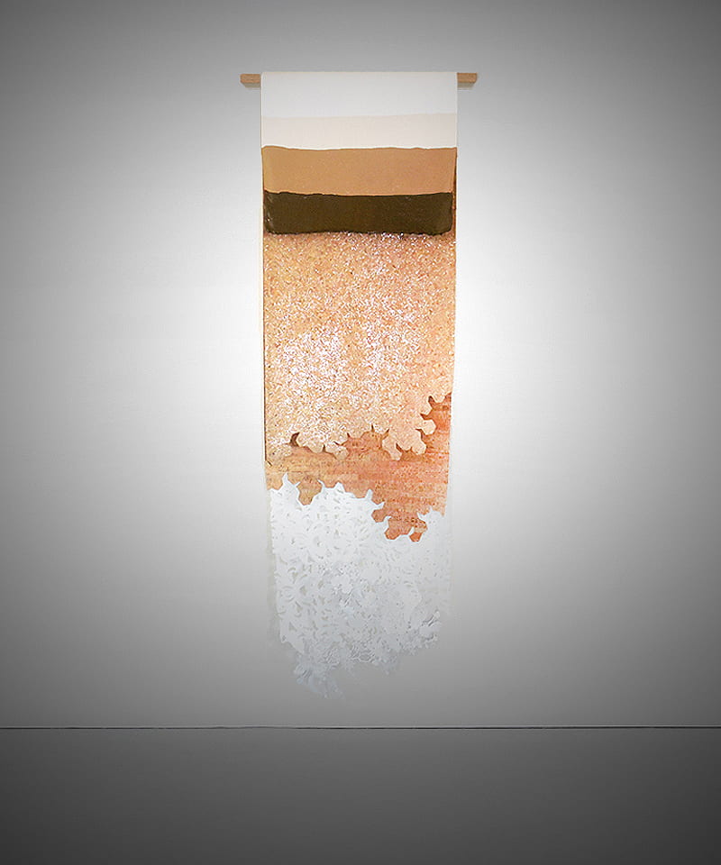
Mystical Artifacts Beneath a Microscope
Digital print on plexiglass / charcoal + pastel + acrylic on glassine / metallic + cotton thread on silk, 72″ x 24″
by Lisa Lesniak
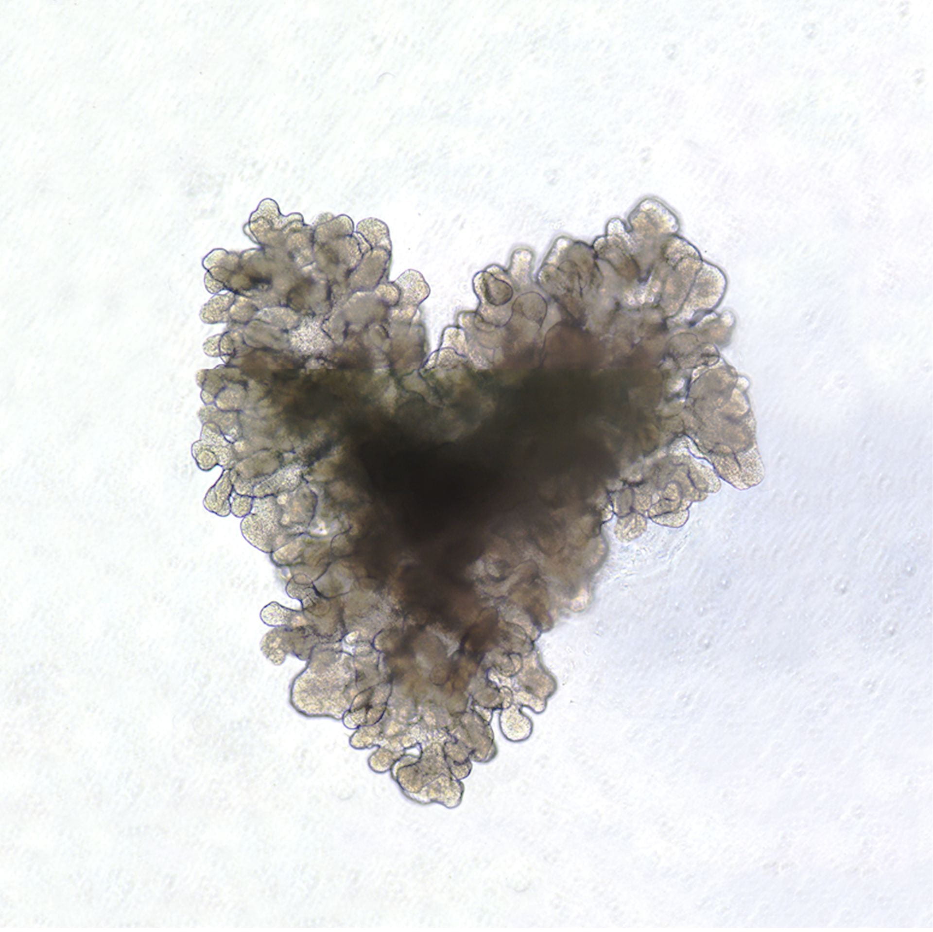

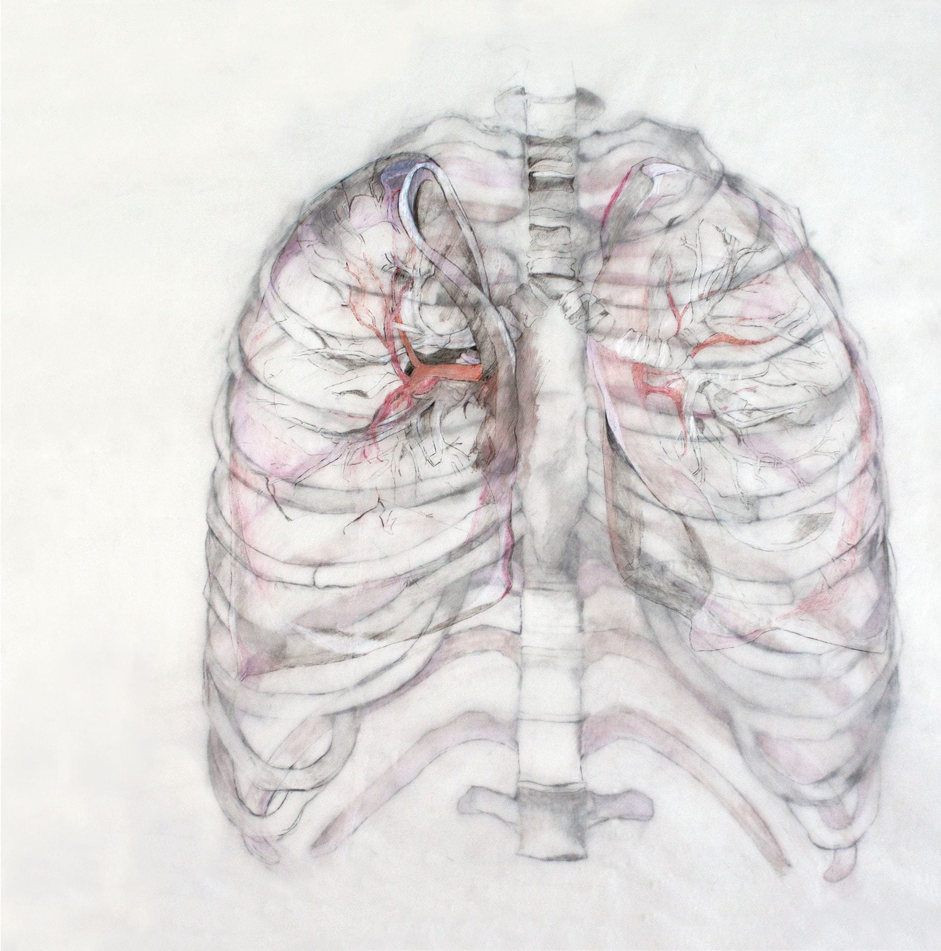
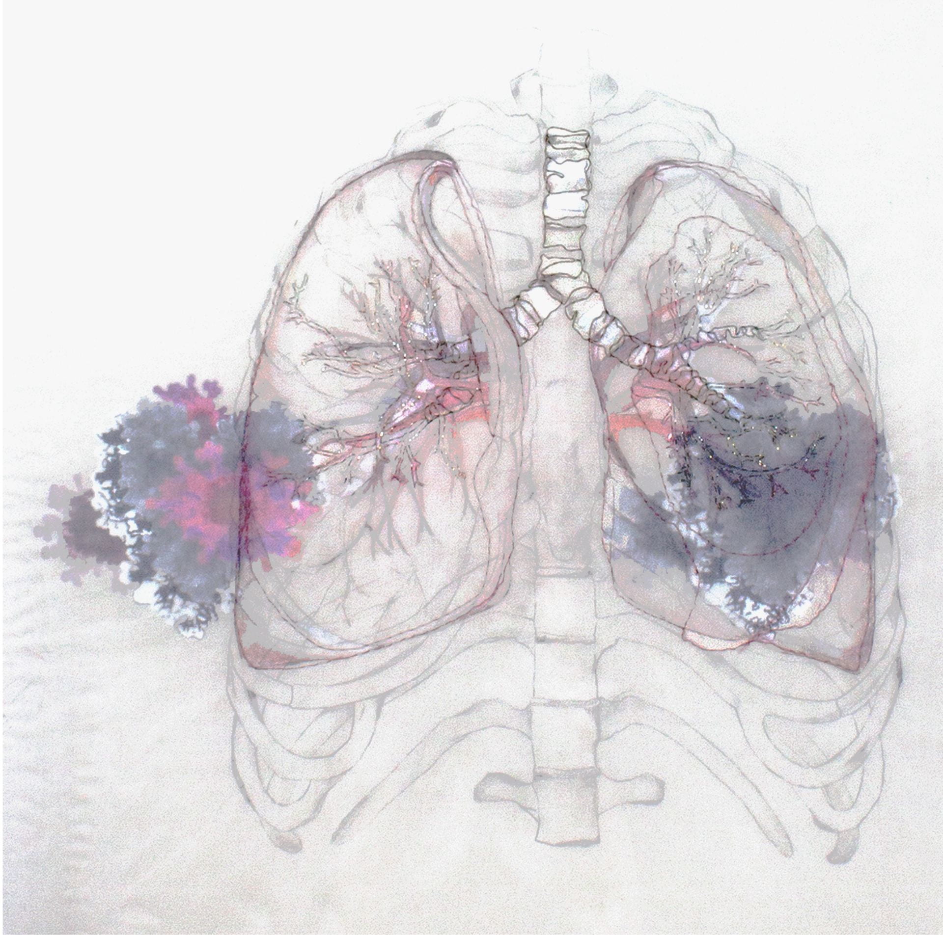
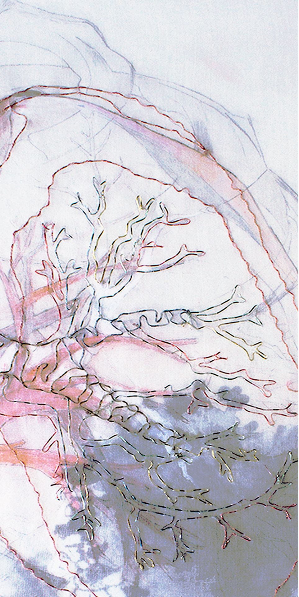
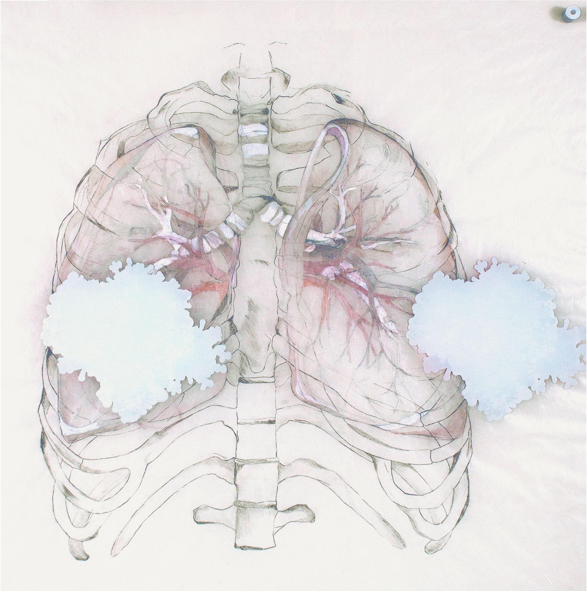
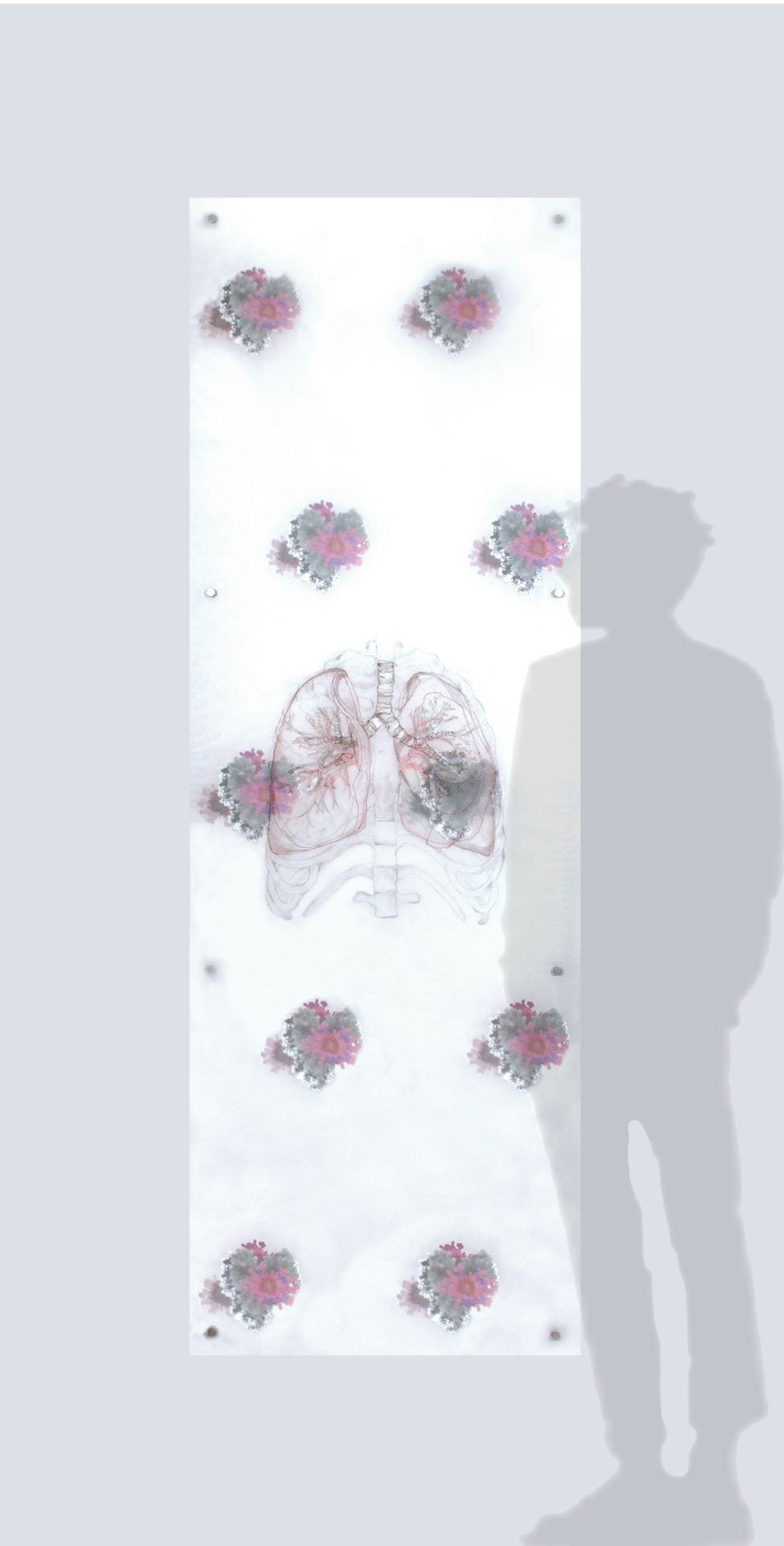
I met with Professor Ya-Wen Chen to discuss the work conducted in her laboratory. While her presentation was clearly informative, the daily workings of the lab remain a mystery. That opacity drove my artistic process. After the first of our two meetings, Professor Chen emailed me a photo of a lung organoid that had grown into a heart shape on February 14th. Both lung and heart, I began to grow and multiply this sample onto an imaginary wall. Layered upon these repeated cell motifs are life-size drawings of lungs and their structural protector, the ribcage. Glassine, a semitransparent paper used between pages to shield prints or illustrations, provides the surface for this double-sided drawing. Placed above, metallic strands trace contours of the lungs, trachea, bronchial tubes, arteries and veins and are sutured to a “pleural” silk membrane. From cell to anatomy to breath, each surface is compressed between two layers of acrylic not unlike a microscopic slide, specimen and its coverslip. Informed by Cubism where all sides of an object are flattened against a picture plane, this act of compression and translucency points to a longtime interest in drawing as seeing through to a transcendent other (side). For this virtual exhibition, the piece 72” x 24” is displayed “in-situ” and is presented along with layer details. Included are close ups of both obverse and reverse.
Email: lesniak.studio@gmail.com
Website: https://lisalesniak.wordpress.com
Instagram: @lillepin
Our lab focuses on using human pluripotent stem cells, including embryonic stem cells and induced pluripotent stem cells, to study human lung development, model lung diseases, lung injury, stem cell-based therapy, and regenerative medicine. Another interest of the lab is to reconstruct the lung via bioengineering approaches. To do so, we have developed a lung organoid, mini lungs in a dish, model that is able to recapitulate human lung development, including generating branching morphogenesis and alveolarization in a dish. We are investigating the potentials of these lung organoids to repair injured lungs and rescue disqualified donor lungs.