
Richard Judelson, MD, Wafaa Elatre, MD, MPH
Clinical History
59 year-old female who was recently diagnosed with solid papillary carcinoma of the left breast and is status post lumpectomy, adjuvant radiation and hormone therapy. Surveillance MRI identified a 2.9 cm left supraclavicular mass just below the thyroid gland, which was suspicious for lymphadenopathy vs a thyroid or parathyroid nodule. Follow up ultrasound of the head & neck confirmed an ovoid shaped, solid mass inferior to the thyroid gland which appears to be contiguous with the left thyroid and devoid of calcifications. This mass measures 2.4 cm and is consistent with TIRADS 4. The patient subsequently underwent ultrasound-guided fine needle aspiration of the neck mass.
Cytomorphologic findings
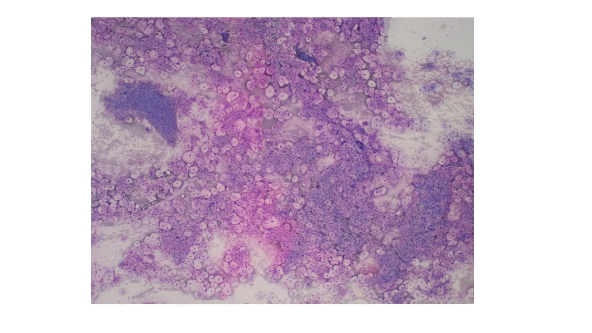
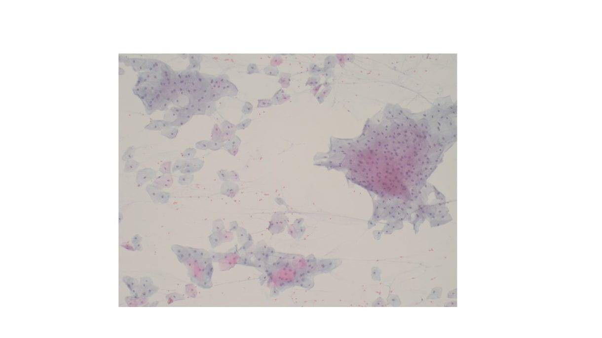
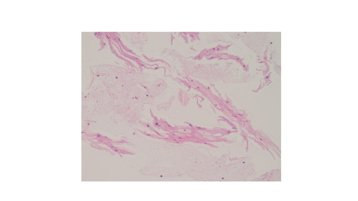
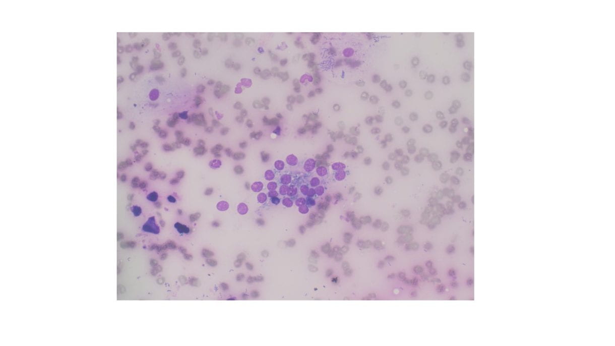
Cellular Diff-Quik and Papanicolaou stained smears showed abundant mature squamous cells with associated bacterial organisms. Rare bland follicular cells admixed with watery and thick colloid and mixed inflammatory cells were present in the background. The cell block showed mature squamous cells, bacterial organisms and mixed inflammation.

Discussion
Suspected thyroid nodules consisting almost entirely of mature squamous epithelial cells is rare. The differential diagnosis of benign appearing squamous epithelium in the thyroid includes squamous metaplasia, thyroglossal duct cyst, thymic/lymphoepithelial/ultimobranchial body remnants, epidermoid/dermoid cysts or esophageal tissue (accidental esophageal sampling vs esophageal diverticula). Any squamous atypia should raise suspicion of a malignant entity such as papillary thyroid carcinoma with squamous changes or a metastatic squamous cell carcinoma.
Differentiating between the various benign squamous lined cystic lesions in and around the thyroid is challenging and definitive diagnosis is best reserved for excisional biopsy.
Of note, the presence of mostly bland, mature squamous epithelium associated bacteria and mixed inflammation is highly suspicious for sampling of esophageal mucosa. In such a case, recommending radiologic follow up with barium studies may be helpful to rule out the possibility of an esophageal diverticula.
Reference
Chen AL, Renshaw AA, Faquin WC, Alexander EK, Heller HT, Cibas ES. Thyroid FNA biopsies comprised of abundant, mature squamous cells can be reported as benign: A cytologic study of 18 patients with clinical correlation. Cancer Cytopathol. 2018 May;126(5):336-341. doi: 10.1002/cncy.21976. Epub 2018 Apr 10. PMID: 29634853.