ATYPICAL MENINGIOMA (WHO GRADE II) CHRISTINE J. SALIBAY, DO ARIANNETTE ROBLED-GOMEZ, MD, WAFAA ELATRE, MD, MPH

64 year-old male with past medical history of meningioma resected in 1998 with recurrence and re-resection in 2006 who presented with a chief of complaint of worsening left sided nasal congestion, difficulty breathing for two weeks and a new right neck mass.  Physical exam showed a 4 to 5 cm well-circumscribed mobile, nontender mass in the right lateral neck without overlying skin changes.

Computed Tomography (CT) imaging of the paranasal sinuses was performed and showed a multi-compartmental and transpatial mass.  The right maxillary sinus/posterior nasal cavity component measured approximately 5.6 x 5.6 x 3.9 cm, extended across midline, and abutted the left inferior turbinate.  The second component centered in the right parapharyngeal, parotid and masticator spaces and measured approximately 5.9 x 3.2 x 2.8 cm.  The third largest component was centered in the right parasellar region and extended into the right sphenoid sinus, right cavernous sinus, Meckel cavity and right orbital apex and measured approximately 2.8 x 2.7 x 1.0 cm.


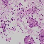
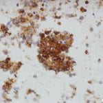
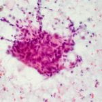
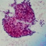
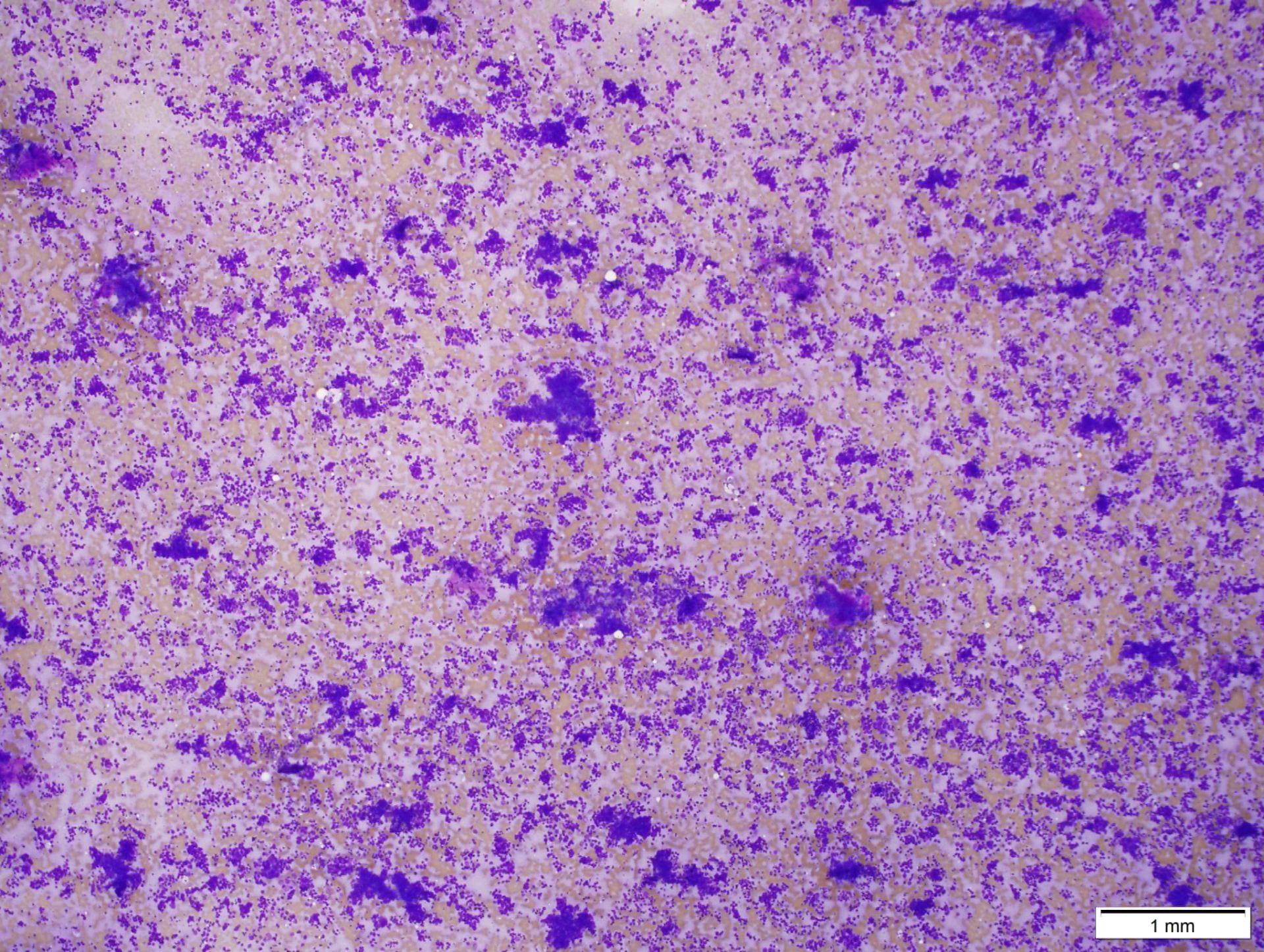
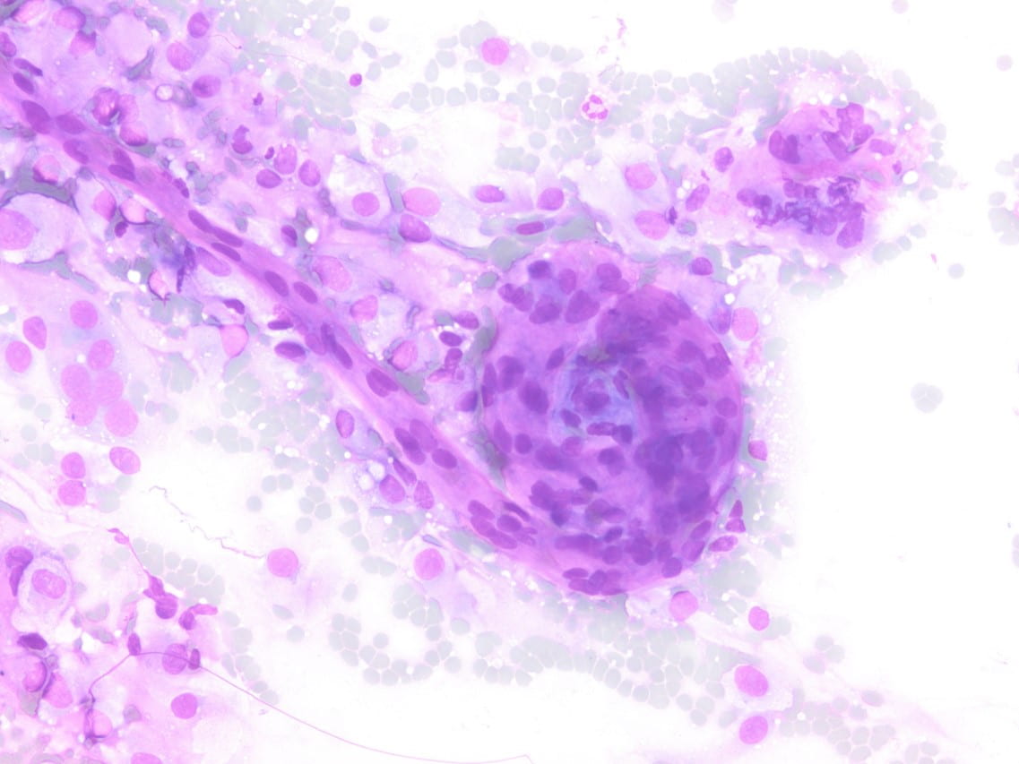
Figure 2. Diff-quick preparation shows whorling architecture (200X).

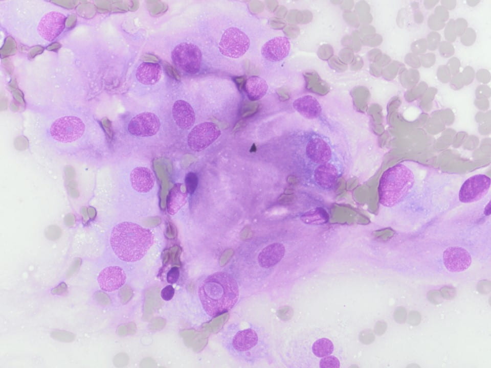
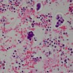
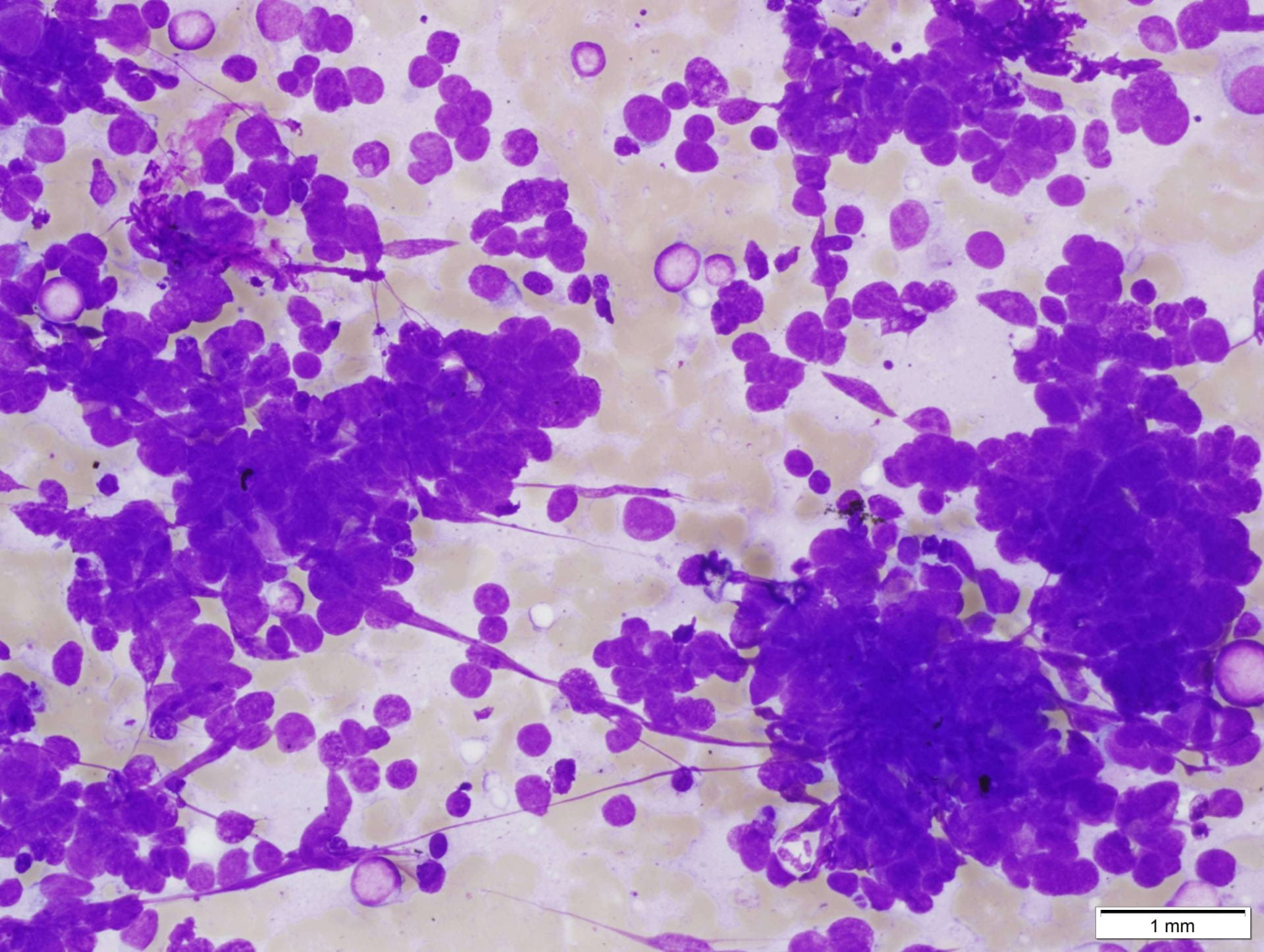
Figure 3. Upon closer inspection, Diff-quick preparations show a) nuclear inclusions (200X) and b) occasional mitotic figures [highlighted by arrow] (200X).
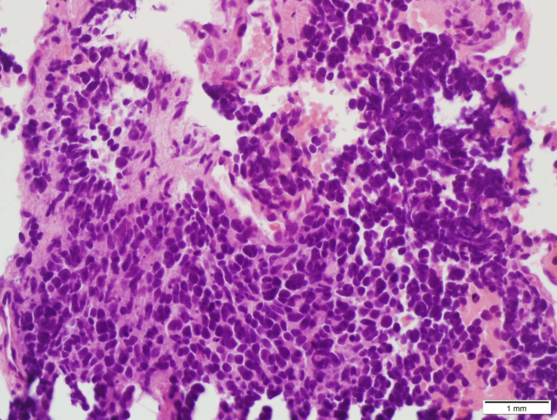
H&E SLIDE:

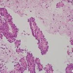
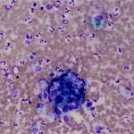
Figure 4. Hematoxylin and eosin (H&E) stain shows a) patternless or sheet-like growth of syncytial cells with areas of increased cellularity (100X) and b) areas of necrosis (200X).  High-power views highlight c) prominent nucleoli and a mitotic figure (400X) with d) nuclear inclusions (200X).

ANCILLARY STUDIES:

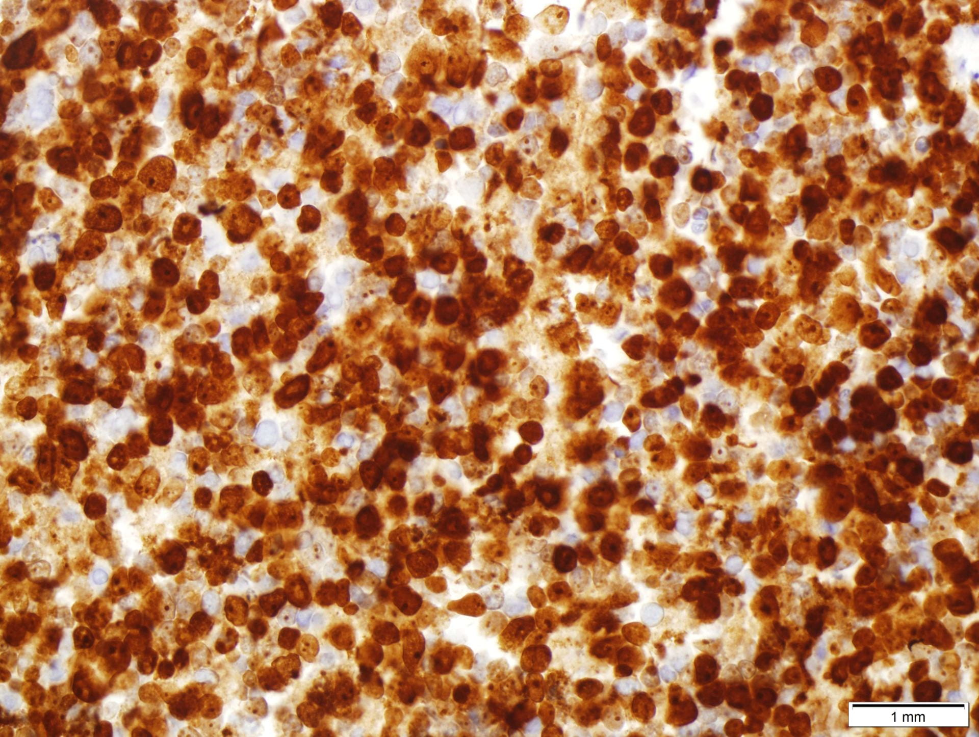
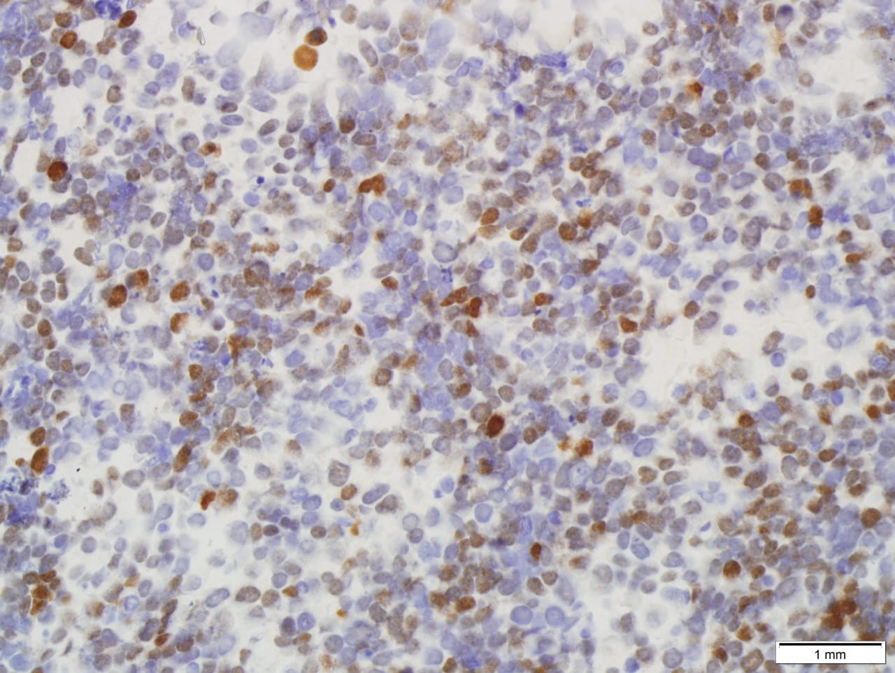
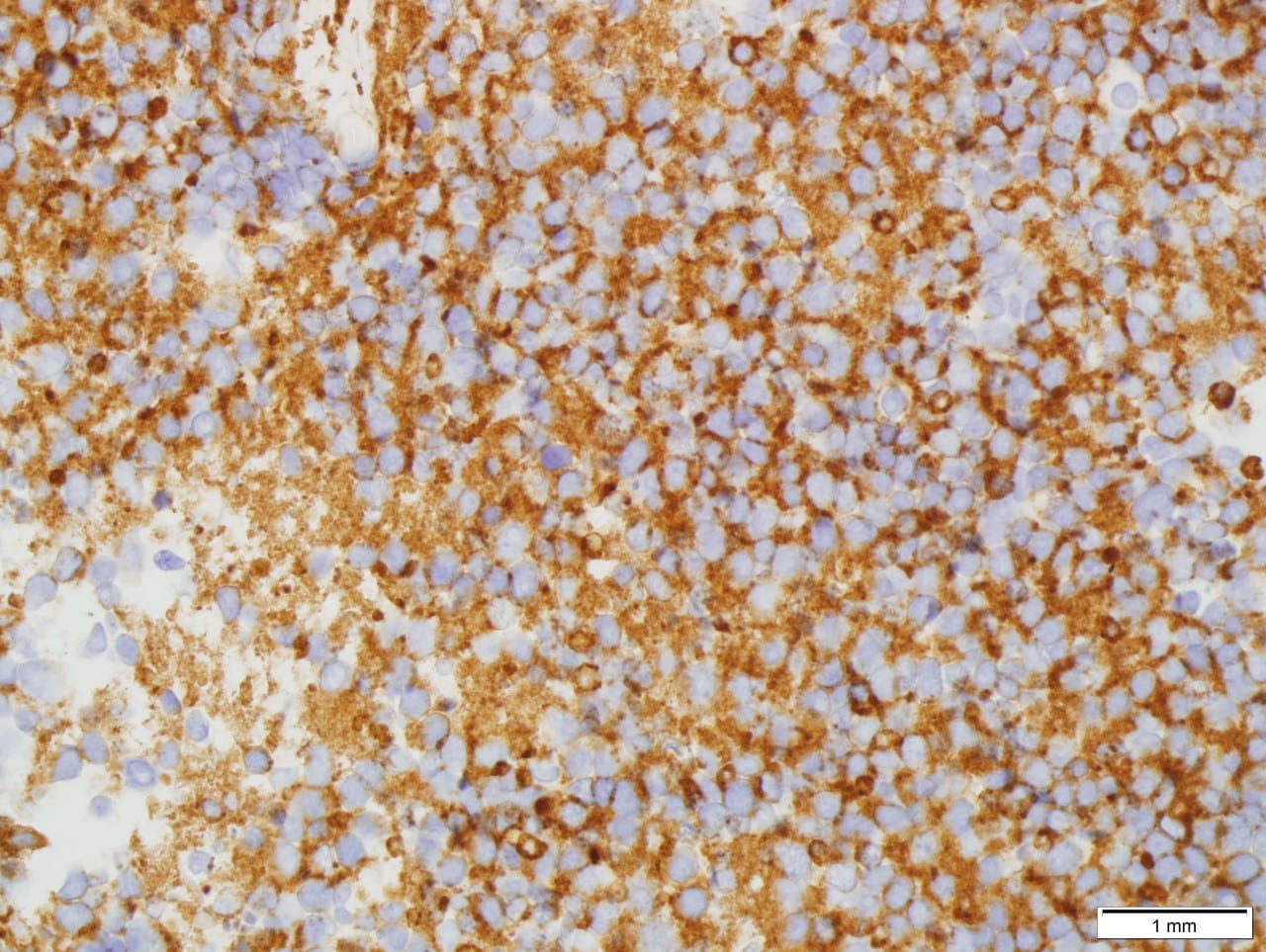
Immunohistochemical stains performed showed the following results:

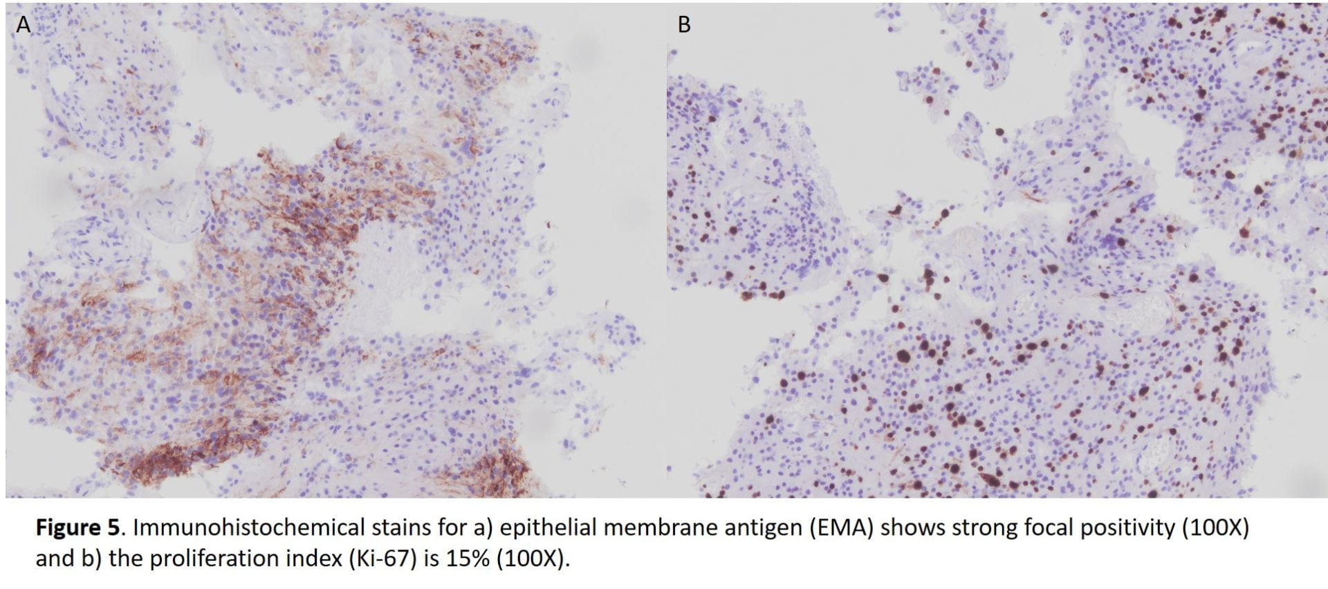
All immunohistochemical stains performed are summarized in the following table.

Table 1. Summary of immunohistochemical stains.

Immunostain Results
EMA Focally positive
Progesterone receptor (PR) Negative
CK AE1/AE3 Negative
S100 Negative
Ki-67 15%


DISCUSSION:

Meningiomas are the most common primary central nervous system tumor believed to arise from the arachnoidal (meningothelial cell) [1,2]. They occur in middle-aged to elderly patients with a peak incidence in the sixth decade with a predilection for female gender [1,2].  Although often sporadic, multiple meningiomas can be associated with a hereditary predisposition such as neurofibromatosis type 2 [1,2].   Intracranial meningiomas occur in cerebral convexities and less likely the intraventricular region and orbits [1,2]. Magnetic resonance imaging shows a dural mass with contrast enhancement and characteristic “dural tail” [1,2].  Clinically, they are generally slow growing and benign with neurologic symptoms arising due to compressive behavior; there are infrequent cases of recurrence associated with increased mortality [1,2].


Recurrence is associated with higher histologic grade and extent of surgical excision [1,2,3].  Increasing grade is associated with increased recurrence with a recurrence risk of 50-55% for grade 2 meningiomas [1,2,3].   The WHO 2016 describes 16 histologic subtypes with three grades [1,2,3]. Grade II meningiomas include three different histotypes, i.e. atypical, chordoid and clear cell [2,3].  Histologic criteria for grade II meningiomas includes (1) three or more of minor criteria: increased cellularity, small cells with a high nuclear/cytoplasmic ratio, prominent nucleoli (readily visible at 10x), uninterrupted patternless or sheet-like growth, and foci of spontaneous or geographic necrosis; or (2) increased mitotic figures, defined as 4 or more mitoses per 10 high-power fields (HPF) or brain invasion [1,2,3].  As in our case, the patient reaches at least three of the minor criteria with increased cellularity, sheet-like growth, prominent nucleoli, and small cell change.  The necrosis identified on the case most likely signifies therapy-related changes.  Mitotic figures although observed in our case do not reach the 4 mitotic figure per 10 hpf threshold, possibly due to insufficient sampling of the tumor.


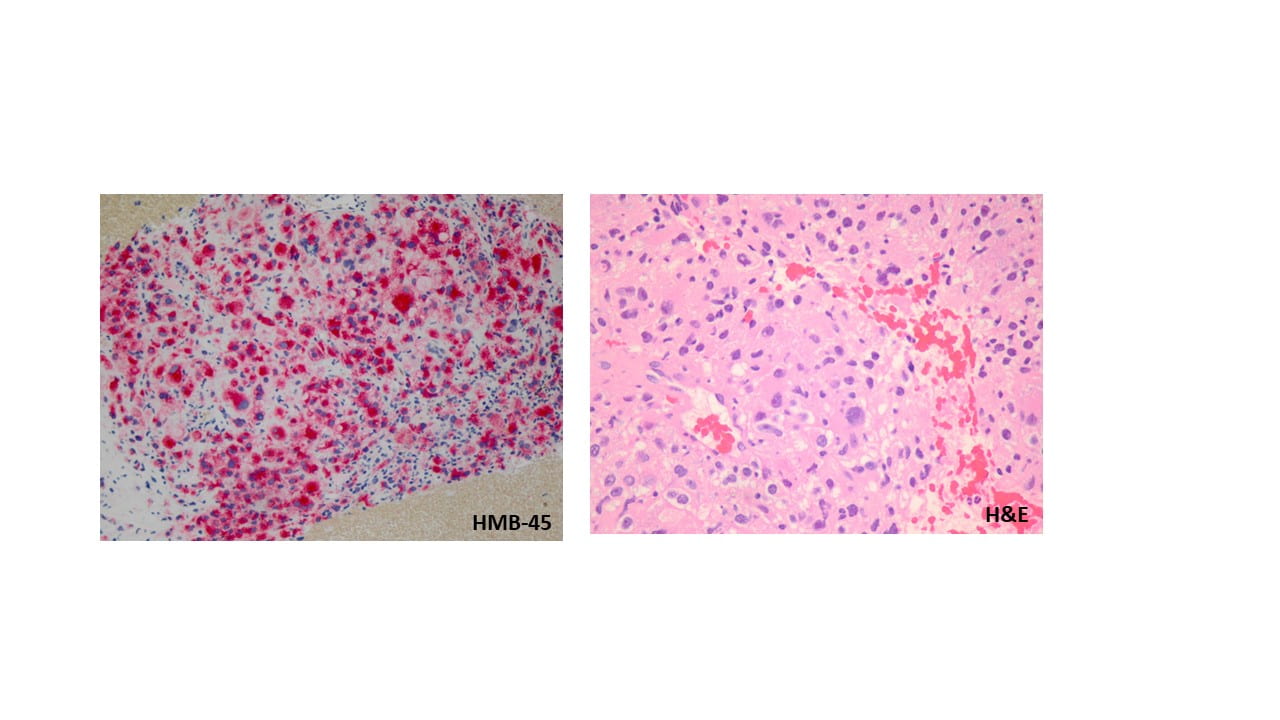
Cytologic features of meningiomas include syncytial clusters of spindled cells arranged in lobules with chromatin clearing and wispy cytoplasm [1,4].  Cells may be arranged in “whorls” that are characteristic of a meningioma, albeit typically low-grade [1,4].  Psammoma bodies and nuclear pseudoinclusions may be seen [4]. Abundant bright eosinophilic psammoma bodies may be seen in secretory meningioma [4].  Degenerative changes may be associated with cells with atypical nuclei with smudgy chromatin and abundant cytoplasm [4].  Atypical meningiomas are associated with increased cellularity and sheet-like growth, as observed with our case [1,4,5].  If there is an abundant necrotic background, one should consider preoperative embolization therapy or other etiologies such as metastatic carcinoma [4].  On H&E, meningiomas show similar features to cytology such as syncytial cells with whorling architecture and clear chromatin patterns; psammoma bodies and nuclear inclusions may be present.  A differential diagnosis may include metastatic carcinoma, solitary fibrous tumor, and hemangiopericytomas; all which lack the characteristic whorls or delicate cytoplasm seen in meningiomas [4].


Meningiomas show positive immunohistochemistry for epithelial membrane antigen (EMA); a more recent immunostain, somatostatin receptor 2A (SST2A), has also shown to be positivity in recent studies [6].  70-80% of meningiomas are also positive for progesterone receptor and lesser immunostaining with estrogen receptor [7,8].  In some studies, male gender, as observe in our case, serves as a negative prognostic factor and a correlation between meningiomas that are less likely to stain for progesterone  receptor are associated with malignant meningioma [2,3,5].  This progesterone receptor-negative immunophenotype is seen in our case.  A Ki-67 proliferation index over 4%, also observed in our case, has also been correlated with increased recurrence risk, but is most commonly used as an adjunct to standard WHO grading and not as an independent factor [1,2].


Additionally, brain invasion and a mitotic rate have been shown to be the most important prognostic indicators of recurrence in meningiomas, especially after gross total resection [3,5]. Minor criteria of hypercellularity, sheeting, macronucleoli, and small cell change is associated with decreased progression free survival [5].


Awareness of this common central nervous system tumor and its histologic grading, specifically in the context of cytopathology, is of utmost importance for all pathologists.


REFERENCES:

  1. Perry A, Louis DN, Budka H et al (2016) Meningiomas. In: Louis DN, Ohgaki H, Wiestler OD, Cavenee WK, Ellison DW, Figarella- Branger D, Perry A, Refeinberger G, von Deimling A (eds) WHO classification of tumours of the central nervous system. IARC press, Lyon, pp 232–245.
  2. Buerki RA, Horbinski CM, Kruser T, Horowitz PM, James CD, Lukas RV. An overview of meningiomas. Future Oncol. 2018;14(21):2161-2177.
  3. Barresi V, Lionti S, Caliri S, Caffo M. Histopathological features to define atypical meningioma: What does really matter for prognosis? Brain Tumor Pathol. 2018;35(3):168-180.
  4. Field, AS and Zarka, MA. Practical cytopathology: A diagnostic approach to fine needle aspiration biopsy. Elsevier; 2017: 483-533.
  5. Perry, Arie, Stafford, Scott, Scheithauer, Bernd, Suman, Vera, Lohse, Christine. Meningioma Grading: An Analysis of Histologic Parameters. Am J Surg Pathol. 1997;21(12):1455-1465.
  6. Menke JR, Raleigh DR, Gown AM, Thomas S, Perry A, Tihan T. Somatostatin receptor 2a is a more sensitive diagnostic marker of meningioma than epithelial membrane antigen. Acta Neuropathologica 130(3), 441–443 (2015).
  7. Hsu DW, Efird JT, Hedley-Whyte ET. Progesterone and estrogen receptors in meningiomas: prognostic considerations. Neurosurg. 86(1), 113–120 (1997).
  8. Pravdenkova S, Al-Mefty O, Sawyer J, Husain M. Progesterone and estrogen receptors: opposing prognostic indicators in meningiomas. Neurosurg. 105(2), 163–173 (2006).


Small Cell Carcinoma of Lung Wenjing Shi, MD, Wafaa Elatre, MD, MPH

63-year-old male with history of 30-pack-year of smoking presented with  cough. The chest CT showed 3.9 cm left upper lobe mass centered near the left hilum with extension into the left mainstem bronchus. Bilateral mediastinal lymphadenopathy and left supraclavicular lymph nodes enlargement were seen.

EBUS-guided FNA of level 4R lymph node was performed.

On Diff-Quik preparation, low power (4x) view is hypercellular. The tumor cells are loosely cohesive or forming small clusters. At high power (40x) magnification, the tumor cells are about 2 times the size of lymphocytes. They have high n/c ratio, very scant cytoplasm, round to oval nuclei, relatively smooth nuclear membrane, and inconspicuous nucleoli. Some cells show molding and crush artifacts. Abundant apoptotic bodies can be readily appreciated. Background of lymphocytes are present.

On Pap stained slides, at high power (40x) magnification, the tumor cells again are small (2 times the size of lymphocytes). They are either loose cohesive or aggregated into clusters with overlapping, molding and crush artifact. The cells have minimal cytoplasm, but round to oval and hyperchromatic nuclei, granular “salt-and-pepper” chromatin, and inconspicuous nucleoli. Mitosis and many apoptotic cells are readily seen. Lymphocytes are present in the background.

Cell block is hypercellular with marked crush artifacts. The small to medium sized, round to oval and hyperchromatic tumor cells show high n/c ratio, minimal cytoplasm, molding and smudging nuclei with finely dispersed chromatin, lack of distinct nucleoli. Apoptosis and mitosis are seen. Immunohistochemically, the tumor cells are moderately to strongly positive for TTF-1, strongly and diffusely positive for synaptophysin. Mitotic index of Ki67 is very high (greater than 90%).


Diff Quick stained slide
Diff quick stained slide high power
cell block
KI67
TTF-1
synaptophysin

Discussion: Pulmonary small cell carcinoma is a high-grade neuroendocrine carcinoma, accounting for 20-25% of all primary lung carcinomas. It is thought to be derived from Kulchitsky cells. Most patients are male smoker (80%). Patients usually present with rapid growing and metastasizing lung mass. Instead of surgical resection, the main treatment for small cell carcinoma is chemotherapy and radiation. The prognosis is very poor with months of the life expectancy

Principle cytologic features of small cell carcinoma include that the tumor cells average approximately 1.5-2 times the size of lymphocytes with scant cytoplasm, finely granular chromatin with a typical salt-and-pepper granular appearance, absent to inconspicuous nucleoli, nuclear molding, and associated apoptosis and mitotic figures. Immunohistochemically, the tumor cells show neuroendocrine markers including CD56, synaptophysin, chromogranin and INSM. TTF-1 is typically positive. Due to the minimal cytoplasm, pan-cytokeratin usually weak/focal or even negative.

Distinguishing between small cell carcinoma from poorly differentiated non-small cell carcinoma and lymphoma is important because of significant differences in patients’ management and prognosis. Lymphoid cells are typically discohesive or singly dispersed without forming cell clusters. Background of lymphograndular bodies is a helpful clue for lymphoid cells.  Small cell carcinoma sometimes is difficult to distinguish from non-small cell carcinoma, particularly squamous cell carcinoma with basaloid features and adenocarcinoma. Finely granular chromatin, inconspicuous nucleoli, nuclear molding and crush artifact are useful features for small cell carcinoma. In difficulty cases, immunohistochemical panels include p40, neuroendocrine markers, Napsin A and TTF-1 will help.

References:

Cibas, ES; Ducatman, BS. Cytology: Diagnostic principle and clinical correlation (4th ed.)

Malignant Perivascular Epithelioid Cell Tumor of the Lung (PECOMA): Catherine Suen, MD, Wafaa Elatre, MD

Clinical information:

55-year-old male with history of asthma and reported history of previous lung granuloma presented to an outside hospital with persistent cough for 9 months and was found to have a 10 x 10 x 10 cm left lower lobe mass on imaging. PET CT showed the large lung mass with hypermetabolic activity, but without evidence of hypermetabolic activity elsewhere. CT guided biopsies were performed at an outside hospital which were reported to have poorly differentiated malignant cells and necrosis, but were inconclusive for a specific pathologic diagnosis. The case was sent to Mayo Clinic for consultation and was reported to most likely represent a poorly differentiated non-small cell carcinoma. This previous biopsy material is not available to us for review. An EBUS with FNA biopsy was performed at Keck Hospital.

Microscopic findings, cytologic features, and immunohistochemical profile:

Moderately cellular smears show small clusters of and singly dispersed large atypical cells with marked anisonucleosis and poikilocytosis, round to oval nuclei, clumped coarse chromatin, multiple prominent nucleoli, and abundant foamy to granular cytoplasm. Numerous multinucleated giant cells are seen. The background consists of blood and blood elements. The cell block shows similar findings with the atypical cells growing around thin-walled capillary vessels and foci showing intranuclear pseudoinclusions. Mitoses and necrosis is not identified.

The tumor cells are positive for HMB45, patchy positive for Melan-A, SMA and TFE3 while negative for S100, SOX10, and AE1/AE3. The KI-67 proliferative index was about 5%.

Of note, the concurrent lymph nodes sampled were all negative for malignant cells.

Discussion:

Perivascular Epithelioid Cell tumors (PEComa) are a distinctive class of mesenchymal neoplasms arising from perivascular epithelioid cells demonstrating combined myomelanocytic markers. They are rare and have a marked female predominance. PEComas of the lung are usually benign and include defined subtypes such as angiomyolipoma (AML), clear cell sugar tumor, and lymphangioleiomyomatosis (LAM). These tumors frequently show association with tuberous sclerosis complex (TSC) and have frequent deletions of the TSC2 gene. Most others are sporadic and a small subset are associated with TFE3 gene fusions.

Our case showed overexpression of TFE3 immunohistochemical stain, which is seen in tumors associated with TFE3 gene fusions. This may have therapeutic implications for aggressive tumor in the future.

Malignant lung PEComas are exceedingly rare, with about 8 reported cases in the literature. Primary diagnostic criteria for malignant PEComa include: prominent coagulative necrosis, mitotic figures ≥ 1/50HP, invasion of adjacent pleura or viscera, and evidence of distant metastasis of homology. Secondary diagnostic criteria include: tumor size ≥ 3 cm, spotty necrosis, high mitotic index ≥ 5%, multinucleated tumor giant cell ≥ 5/50HP, marked hypercellularity, nuclear atypia and pleomorphism, and numerous intranuclear pseudoinclusions. Criteria for diagnosis of malignant lung PEComa is such that if the tumor meets 1-2 major diagnostic criteria and/or ≥1 secondary diagnostic criteria. If the tumor satisfies only ≥ 2 secondary diagnostic criteria, the suggested diagnosis is lung PEComa with malignant potential.

As the tumor in this case met many secondary criteria, but there was no evidence of mitosis or necrosis in our limited sample, we deferred the distinction between benign vs malignant to the resection specimen.

References:

Zhao, J. et al. Malignant perivascular epithelioid cell tumor of the lung synchronous with a primary adenocarcinoma: one case report and review of the literature. BMC Cancer (2019) 19:235.

Armah, HB, Parwani, AV. Perivascular Epithelioid Cell Tumor. Archives Pathol Lab Med (2009) 133: 648-654.

Folpe AL. Neoplasms with perivascular epithelioid cell differentiation (PEComas). In: Fletcher CDM, Unni KK, Mertens R, eds. Pathology and Genetics of Tumors of Soft Tissue and Bone. Lyon, France: IARC Press; 2002: 221-222. World Health Organization Classification of Tumors.

Saluia K et al: Malignant perivascular epithelioid cell tumor of the oropharynx with strong TFE3 expression mimicking alveolar soft part sarcoma: a case report and review of the literature. Hum Pathol, 76: 149-55, 2018.

Metastatic Renal Cell Carcinoma: Sonia Veran-Taguibao, MD, Wafaa Elatre, MD

49 year old male with history of hyperlipidemia, hypertension, CKD Stage III and DM presents to the ED with nausea and vomiting along with diffuse constant mild abdominal pain. CT Abdomen Pelvis revealed a left 7.3 cm renal mass, numerous scattered pulmonary nodules and a lesion in the right hepatic lobe (segment 5 and 6). Fine needle aspiration biopsy of the liver revealed metastatic renal cell carcinoma


Definition

  • Malignant neoplasm of the kidney made up of epithelial cells with clear or granular cytoplasm arising from proximal convoluted tubule
  • First described by Daniel Sennert in 1613
  • Introduced by Foot and Humphreys, and Foote et al because of the renal tubular origin of these tumors

Essential features

  • Sixth and seventh decades
  • Risk factors include smoking, obesity and hypertension. Less commonly, inherited (tuberous sclerosis, Birt-Hogg-Dube and VHL syndromes) and sickle cell disease

Terminology

  • Conventional renal cell carcinoma
  • Thought to have ascend from adrenal rest and were called ‘hypernephromas’
  • Nephrocellular carcinoma
  • Grawitz tumor


Epidemiology

  • Male predominance
  • Caucasian>Asian>African American
  • North America, Europe and Australia/New Zealand
  • Approximately 90% of the renal epithelial carcinoma
  • About 70% of all renal cortical malignancies


Sites

  • Renal cortex
  • Upper pole of the kidney
  • Known to metastasize to weird sites like parotid, anus and thyroid
  • Metastasize via hematogenous route


Clinical features

  • Triad of hematuria (most common), abdominal pain and a flank mass


Radiology description

  • Average size: 7 cm
    • Heterogenous appearance due to necrosis, hemorrhage and cystic degenerations
    • Associated with hypervascular retroperitoneal lymphadenopathy


    Prognosis and treatment

    • Prognosis depends on histologic grade, sarcomatoid or rhabdoid differentiation, metastasis and tumor necrosis (>10%)
    • Poor prognosis if there is loss in chromosome 14q, 4p and 9p
    • 5 years survival is 60-80% in Stage I and 5% in Stage IV
    • Arterial embolization
    • Partial or Radical nephrectomy
    • Interleukin 2, sunitinib (Anti-VEGF TKI) therapy, mTOR inhibitor (targeted) therapy


    Cytology description

    • Clusters of cohesive cells with large round slightly eccentrical placed nucleus and abundant wispy cytoplasm with ill defined edges

    Positive stains

    • CD10
    • RCC
    • CA-IX
    • PAX 8
    • PAX 2
    • EMA

    Negative stains

    • CK 7
    • CK20
    • Hale’s colloidal iron
    • Inhibin
    • CD68


    Molecular/cytogenetics description

    • Somatic mutations or deletions of hypermethylation of chromosome 3p
    • Non random chromosomal changes on chromosomes 3p and 14q
    • Gain of chromosome 5q22-qter and trisomy 7

    Differential diagnosis

    • Histocytes
    • Benign tubular cells
    • Angiomyolipoma
    • Adrenal cortical cells

Carcinoid Tumor of the Lung: Tom Liang, MD, Robert Weller, Wafaa Elatre, MD

  • 57 year old male with a past medical history of hypertension, type 2 diabetes, GERD, and asthma presented with 2 months of hemoptysis, weight loss, night sweats.
  • Reports a history of positive PPD, but has never been treated for tuberculosis. Sputum cultures were AFB-negative x3.
  • Imaging showed a partially calcified left hilar mass with extension and occlusion of the left lower lobe
  • Patient underwent endobronchial ultrasound for further characterization of the mass and FNA sampling

Microscopic findings, architecture & cytologic features:

Cellular smear preparations show numerous singly dispersed and loosely cohesive flat sheets of small atypical cells with high nuclear to cytoplasmic ratios, round to oval hyperchromatic nuclei, salt and pepper chromatin, rare mitotic activity (<2 per high power field), and scant granular cytoplasm in a background of numerous mixed inflammatory cells, ciliated bronchial epithelial cells, and acellular debris. Cell block findings consistent with smear preparations with rosette and nest formations seen.

Final Diagnosis: Carcinoid Tumor


Discussion

Pulmonary neuroendocrine tumors are divided into four classifications: carcinoid, atypical carcinoid, large cell neuroendocrine carcinoma, and small cell carcinoma. Of these entities, typical carcinoid tumors have the best prognosis. Carcinoid tumors are low-grade malignant neoplasms representing 2-3% of pulmonary tumors. Regional lymph nodes may be involved in 10 to 15% of patients and 5 to 10% will eventually have distant metastasis. Patients with typical carcinoid tumors may present with hemoptysis, cough, wheezing, and recurrent pneumonias. Carcinoid syndrome, characterized by flushing, diarrhea, wheezing, and hypotension, rarely occurs. Symptoms depend on the location of the tumor, as peripheral lesions are more likely to be asymptomatic, while central lesions may cause compression of nearby structures and post-obstructive pneumonias. On imaging, peripheral tumors present as a solitary pulmonary nodule, while central lesions may show evidence of compression to the airway and atelectasis. These lesions may also be calcified.


Typical features of carcinoid tumors on cytologic preparations include uniform appearing cells, isolated and in loose clusters. The tumor cells may range from round to oval or elongated and generally have moderate to abundant granular cytoplasm. The nuclei are round with smooth contours and contain “salt and pepper” chromatin. On histologic and cell block sections, the tumor cells may form nest, ribbons, papillae, or rosettes. The typical cytologic features and formations seen on the cell block sections are are consistent with those seen in the case.


The differential diagnosis of carcinoid tumors includes benign bronchial epithelial cells, adenocarcinoma, lymphoma, and other neuroendocrine tumors including atypical carcinoid tumor and small cell carcinoma. Carcinoid tumor cells may appear similar to benign bronchial cells as they have a bland appearance that mimics normal cells, but they will not be ciliated. They may be mistaken for adenocarcinoma due to their propensity to form rosette structures. Carcinoid tumor cells may be distinguished from lymphoid cells by their more abundant cytoplasm. The different pulmonary neuroendocrine tumors must be distinguished from each other by the number of mitotic figures per high-powered field. Typical carcinoid tumors have <2/hpf, atypical carcinoid tumors have 2-10/hpf, and large cell neuroendocrine carcinomas and small cell carcinomas will each have >10/hpf. Immunohistochemical stains are also important in narrowing the differential diagnosis. Chromogranin and synaptophysin stains can readily distinguish tumor cells of neuroendocrine origin and are important in making the diagnosis of carcinoid tumor. Most carcinoid tumors are also positive for keratins, as in this case. TTF-1 may be positive in about a third of carcinoid tumors but was negative in the case. The most important stain in distinguishing carcinoid tumor from other neuroendocrine tumors is Ki-67, a marker for mitotic activity. Less than 25% of cells will stain positive in the less aggressive carcinoid and atypical carcinoid tumors, whereas greater than 50% staining is expected in large cell neuroendocrine carcinomas and small cell carcinomas. A more definitive diagnosis may be made after evaluating histologic features of the resected tumor.


The prognosis for typical carcinoid tumor is excellent because metastasis is rare and slow to occur. Surgical resection of the tumor leads to 5-year survival rates of over 90 percent. The patient in this case has elected for surgical resection of his tumor and is scheduled for surgery.


References:


Chesnutt AN, Chesnutt MS, Prendergast NT, Prendergast TJ. Pulmonary Disorders. In: Papadakis MA, McPhee SJ, Rabow MW. eds. Current Medical Diagnosis & Treatment 2019 New York, NY: McGraw-Hill; http://accessmedicine.mhmedical.com.libproxy1.usc.edu/content.aspx?bookid=2449&sectionid=194434470. Accessed September 19, 2019.


Cibas ES, Ducatman BS. Cytology: Diagnostic Principles and Clinical Correlates. Fourth Edition. Philadelphia, PA. Elselvier Saunders. 2014.


Hendifar, Andrew E., Alberto M. Marchevsky, and Richard Tuli. “Neuroendocrine tumors of the lung: current challenges and advances in the diagnosis and management of well-differentiated disease.” Journal of Thoracic Oncology 12.3 (2017): 425-436.


Okike, Nsidinanya, Philip E. Bernatz, and Lewis B. Woolner. “Carcinoid tumors of the lung.” The Annals of thoracic surgery 22.3 (1976): 270-277.


Solid Pseudopapillary Neoplasm of the Pancreas: Arjun Mehta, MD, Wafaa Elatre, MD

23 year old female who presented to the ED with abdominal pain was found to have a 9.0 cm mass in the body and tail of pancreas without evidence of ductal obstruction or involvement. Attached are the photomicrographs


PAP stained images show numerous vascular channels with tumor cells “budding” off the capillaries. The individual cells display eccentrically placed nuclei and slender cytoplasmic projections.


H&E stained image shows pseudopapillae comprised of capillary cores bordered by tumor cells
 
B- Catenin                            CD10                               PR
IHCs: Beta catenin (nuclear positive) , CD10 (Membranous staining) , Progesterone receptor (Nuclear positive)

Definition

  • Low grade, malignant epithelial tumors with uncertain cellular differentiation
  • First described by V.K Frantz in 1959

Essential features

  • Presents in young women, classically as large body / tail mass
  • Variable amount of solid and cystic formation at gross exam
  • Papillary fronds on myxoid or hyalinized vascular stalk lined by poorly cohesive, uniform cells with nuclear grooves comprising solid and cystic areas
  • β-catenin positive

Terminology

  • Also known as solid pseudopapillary tumor, papillary epithelial neoplasm, papillary cystic neoplasm, solid and papillary neoplasm, low grade papillary neoplasm and Hamoudi or Frantz tumor

Epidemiology

  • Represents 1-2% of pancreatic neoplasms
  • Mainly young females are affected – the female to male ratio is 10:1
  • Usually presents in the third to fourth decade of life (mean age 35 years)
  • In men tends to occur at an older age with more aggressive behavior (Surgery 2008;143:29)

Sites

Clinical features

  • The most common symptoms are abdominal pain and a palpable, non-tender, upper abdominal mass
  • Also symptoms related to an intra-abdominal mass effect, such as discomfort, nausea, vomiting and early satiety

Laboratory

Radiology description

  • Well circumscribed, encapsulated, heterogeneous pancreatic lesion with cystic degeneration on CT or MRI

 Prognostic factors

  • Poor prognostic factors include: size > 5 cm, male gender, necrosis, cellular atypia, vascular invasion, perineural invasion and invasion into adjacent structures (Dig Liver Dis 2013;45:703)

Cytology description

  • Cellular smears with delicate papillary fronds
  • Tumor cells cells are usually bland and uniform with a moderate amount of cytoplasm, which usually contains variable sized clear perinuclear vacuoles or cytoplasmic eosinophilic hyaline globules
  • Nuclei are round to oval with grooves and finely granular chromatin (J Cytol 2010;27:118Arch Pathol Lab Med 2017;141:990)

Positive stains

Negative stains

 Molecular / cytogenetics description

  • Point mutation in exon 3 of β-cateningene (CTNNB1) is present in more than 90%
  • Gene mutation results in the accumulation of β-catenin in the cytoplasm and formation of a β-catenin–Tcf/Lef complex, through which the Wnt signaling pathway activates several oncogenic genes such as MYCand Cyclin D1 (Am J Clin Pathol 2017;149:67)

Scar Endometriosis: Wafaa Elatre, MD

  • 26 year old female
  • Presented with subcutaneous mass (3 x 2 cm) in the right lower abdomen at the edge of cesarean section scar.
  •  She reported cyclic symptoms of pain
  • The patient was then referred for fine needle aspiration cytology (FNAC

  • Cytology FNAC from the abdominal lump was carried out.

  •     

The smears were cellular, consisting of epithelial and stromal fragments. The epithelial cell was arranged in monolayer sheets of polygonal cells with large, hyperchromatic nuclei and moderate amount of cytoplasm, with considerable nuclear overlapping. The stromal aggregates also showed crowded overlapping nuclei and scant admixed hemosiderin-laden macrophages; mild to moderate epithelial atypia was observed.